Figures & data
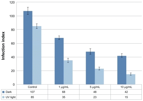
Figure 1 Characterization of silver nanoparticles: (A) transmission electron microscope (TEM) image at the scale of 20 nm, (B) TEM image at the scale of 50 nm, and (C) size distributions with dynamic light scattering analysis.
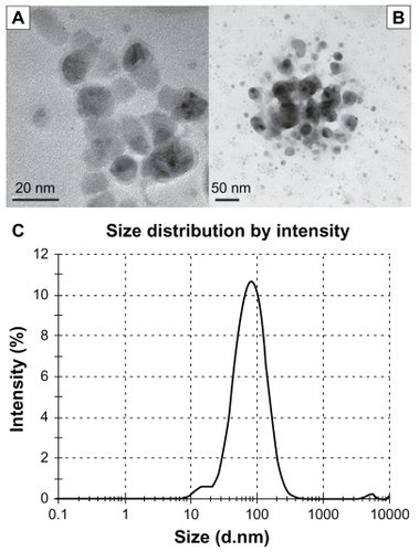
Figure 2 Microscopic view of parasites (A) in the control group (not exposed to silver nanoparticles [Ag-NPs]), (B) in the group exposed to Ag-NPs in the dark, and (C) in the group exposed to Ag-NPs under ultraviolet light (Giemsa staining, magnification 100×).
![Figure 2 Microscopic view of parasites (A) in the control group (not exposed to silver nanoparticles [Ag-NPs]), (B) in the group exposed to Ag-NPs in the dark, and (C) in the group exposed to Ag-NPs under ultraviolet light (Giemsa staining, magnification 100×).](/cms/asset/3dee53db-7ace-404f-af09-30cfbad8e68c/dijn_a_23883_f0002_c.jpg)
Figure 3 Microscopic views of the effects of silver nanoparticles (Ag-NPs) on metabolic activity of Leishmania tropica promastigotes in the dark: (A) parasites in the control group (not exposed to Ag-NPs), (B) the cytopathic effects of Ag-NPs at 150 μg/mL, and (C) the cytopathic effects of Ag-NPs at 200 μg/mL. Arrows in (A) indicate the formation of formazan crystals and in (B) and (C) indicate the clusters of Ag-NPs (magnification 40×).
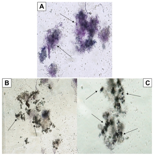
Figure 4 Microscopic views of the effects of silver nanoparticles (Ag-NPs) on metabolic activity of Leishmania parasites under ultraviolet light: (A) parasites in the control group (not exposed to Ag-NPs), (B) the cytopathic effects of Ag-NPs at 150 μg/mL, and (C) the cytopathic effects of Ag-NPs at 200 μg/mL. Arrows in (A) indicate the formation of formazan crystals and in (B) and (C) indicate the clusters of Ag-NPs (magnification 40×).
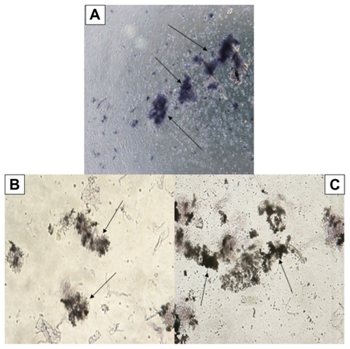
Figure 5 The effects of different concentrations of silver nanoparticles on metabolic activity of Leishmania tropica promastigotes both in the absence and in the presence of ultraviolet (UV) light (I, control group; II, 25 μg/mL; III, 50 μg/mL; IV, 100 μg/mL; V, 150 μg/mL; VI, 200 μg/mL).
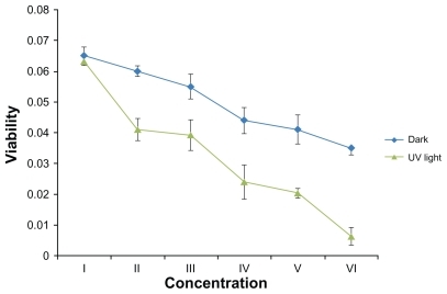
Figure 6 Effects of silver nanoparticles (Ag-NPs) on the proliferation of Leishmania tropica promastigotes in the dark and under ultraviolet (UV) light in different incubation periods (I, parasites in control group in the dark; II, parasites exposed to Ag-NPs in the dark; III, parasites in control group exposed to UV light; IV, parasites exposed to Ag-NPs under UV light). Ag-NPs had significant effects on Leishmania parasites in the experiment groups compared with the control groups. The biggest effect was observed in the experiment group exposed to Ag-NPs and UV light.
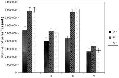
Figure 7 Microscopic views of macrophages infected or not infected by parasites after being stained with Giemsa. Leishmania tropica promastigotes (A) in the control group held in the dark and (B) in the control group exposed to ultraviolet (UV) light infected the macrophages. (C) L. tropica promastigotes exposed to silver nanoparticles (Ag-NPs) in the dark also infected macrophages, but their infectivity was very low compared with the control groups. (D) L. tropica promastigotes exposed to Ag-NPs under UV light did no infect the macrophages. (Arrows indicate macrophage-infected parasites.)
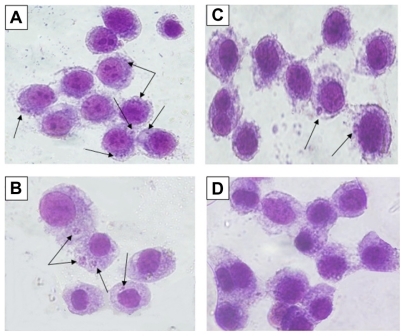
Figure 8 Effects of different concentrations of silver nanoparticles on the infection index of Leishmania tropica amastigotes both in the absence and in the presence of ultraviolet (UV) light.