Figures & data
Figure 1 Size (A) and zeta potential (B) distribution of the FR-targeted liposomes (F-L-Gd/calcein). The mean diameter of the liposomes was 136 nm (A), and the zeta potential was about −13.6 mV (B).
Abbreviation: FR, folate receptor.
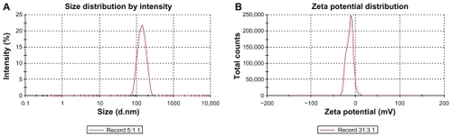
Figure 2 T1-weighted images of Gd-liposomes and Gd-DTPA.
Abbreviations: DTPA, diethylenetriamine pentaacetic acid; Conc., concentration.
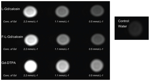
Figure 3 Uptake of liposomal calcein by KB cells. The cells were incubated at 37°C for 1 hour with L-Gd/calcein (A, A′), F-L-Gd/calcein (B, B′), or F-L-Gd/calcein + 1 mM FA (C, C′). Top panels show cells visualized in the phase-contrast mode (A, B, C); bottom panels show the same fields in the fluorescence mode (A′, B′, C′)
Abbreviation: FA, folic acid.
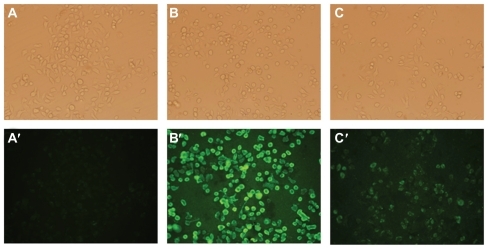
Table 1 Uptake of liposomal calcein by KB, HeLa, and A549 cells determined by flow cytometry
Figure 4 Comparison of the uptake of liposomes between FR-positive (KB, HeLa) and FR-negative (A549) cell lines.
Abbreviations: FR, folate receptor; FA, folic acid.
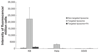
Figure 5 T1-weighted image of HeLa cells after incubated with contrast agents (coronal scan). (A) L. (B) L-Gd/calcein. (C) F-L-Gd/calcein. (D) F-L-Gd/calcein + 1 mM FA. (E) Gd-DTPA.
Abbreviations: FA, folic acid; DTPA, diethylenetriamine pentaacetic acid.
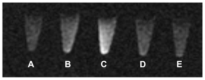
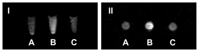
Figure 6 T1-weighted image of KB cells after incubated with contrast agents (I, coronal scan; II, axial scan) (A) L-Gd/calcein. (B) F-L-Gd/calcein. (C) F-L-Gd/calcein + 1 mM FA.
Abbreviation: FA, folic acid.