Figures & data
Table 1 The calculated secondary structure fractions of peptide
Figure 1 After stirring for 48 hours, paclitaxel 1 mg/mL in pure water was still clear (left). However, the same concentration of paclitaxel in 0.5% RADA16 aqueous solution was changed to a colloidal suspension (right).
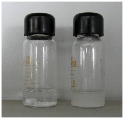
Figure 2 Morphology of RADA16 peptide and RADA16-PTX suspension. (A and C) Nanofibers in RADA16 solution. (B and D) Nanofibers in RADA16- PTX suspension. It can be seen that some PTX particles were coated by RADA16 nanofibers (black arrowhead).
Abbreviation: PTX, paclitaxel.
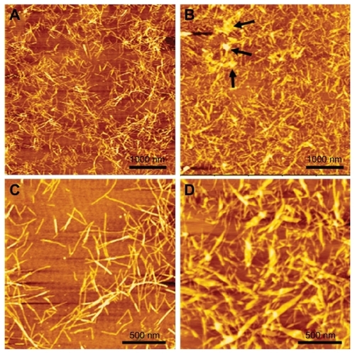
Figure 3 Secondary structure of RADA16 and RADA16 with incorporated paclitaxel. RADA16 peptide had a typical β-sheet structure. However, when paclitaxel was incorporated with RADA16, there was an obvious decrease in β-sheet structure.
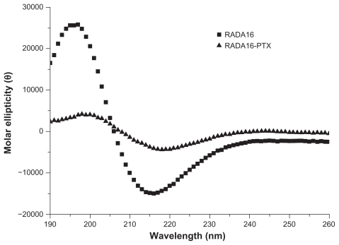
Figure 4 Size distribution of pure RADA16 solution and RADA16-PTX suspension with different peptide concentrations. There was no significant difference between the 0.5% and 1% RADA16-PTX groups.
Abbreviation: PTX, paclitaxel.
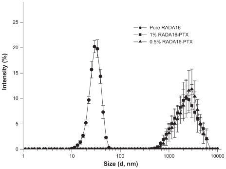
Figure 5 Schematic representation of the interaction between RADA16 peptides and PTX. All alanine residues are present on the hydrophobic face of the RADA16 β-sheet, and the hydrophilic face consists of alternating arginine and aspartic acid residues (carbon, white; oxygen, red; nitrogen, blue; hydrogen, gray).
Abbreviation: PTX, paclitaxel.
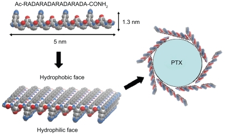
Figure 6 Frequency sweep of RADA16-PTX hydrogel in rheological analysis. (A) 0.5% RADA16-PTX and (B) 1% RADA16-PTX. The results showed that the storage modulus of 0.5% RADA16-PTX hydrogel was much lower than that of 1% RADA16-PTX hydrogel.
Abbreviation: PTX, paclitaxel.
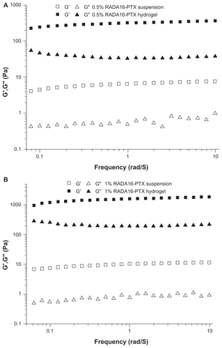
Figure 7 In vitro release of PTX from RADA16-PTX hydrogel with different peptide concentrations. (A) The rates of released PTX at different points and (B) photographs of 1% RADA16-PTX hydrogel in phosphate-buffered saline at 0.5 hours and 48 hours after the start of the release test.
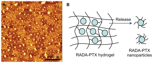
Figure 8 In vitro release of PTX from RADA16-PTX hydrogel. (A) Atomic force microscopy image of supernatant in release medium, in which many nanoparticles could be seen. (B) schematic representation of modeling for PTX release from PTX-RADA16 hydrogel.
Abbreviation: PTX, paclitaxel.
Abbreviation: PTX, paclitaxel.
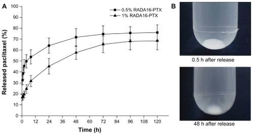
Figure 8 In vitro release of PTX from RADA16-PTX hydrogel. (A) Atomic force microscopy image of supernatant in release medium, in which many nanoparticles could be seen. (B) schematic representation of modeling for PTX release from PTX-RADA16 hydrogel.
Abbreviation: PTX, paclitaxel.
Figure 9 Inhibitory effect of RADA16, PTX and RADA16-PTX hydrogel with different peptide concentrations on the growth of MDA-MB-435S cells in vitro.
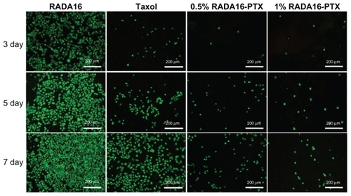
Figure 10 Cell viability assessment by the Live/Dead assay method after treatment with RADA16, PTX, or RADA16-PTX hydrogel with different peptide concentrations.
Abbreviation: PTX, paclitaxel.
Abbreviation: PTX, paclitaxel.
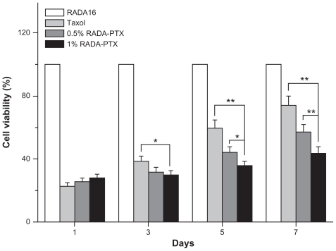
Figure 10 Cell viability assessment by the Live/Dead assay method after treatment with RADA16, PTX, or RADA16-PTX hydrogel with different peptide concentrations.
Abbreviation: PTX, paclitaxel.