Figures & data
Table 1 Serum lipids of rabbits treated with LDE-etoposide (6 mg/kg body weight/week) or saline solution (control group), for 8 weeks
Figure 1 Food intake (A) and bodyweight (B) of rabbits treated with LDE-etoposide or saline solution (control group) over an 8-week period.
Abbreviation: LDE, cholesterol-rich nanoemulsion.
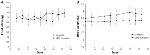
Table 2 Hematological profile of rabbits treated with LDE-etoposide (6 mg/kg body weight/week) or saline solution (control group), for 8 weeks
Table 3 Macroscopic morphometry of aorta in rabbits treated with LDE-etoposide (6 mg/kg body weight/week) or saline solution (control group), for 8 weeks
Figure 2 Aortas of rabbits treated with (A) saline solution (control group) or (B) LDE-etoposide, for 8 weeks, stained by Scarlet Red.
Abbreviation: LDE, cholesterol-rich nanoemulsion.
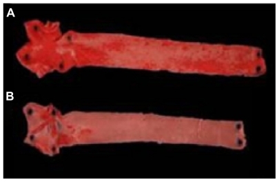
Table 4 Microscopic morphometry (intima and media layers) of aorta of rabbits treated with LDE-etoposide (6 mg/kg body weight/week) or saline solution (control group), for 8 weeks
Table 5 Protein expression (%) of inflammation and proliferation markers and lipoprotein receptors, evaluated by immunohistochemistry of aorta of rabbits treated with LDE-etoposide (6 mg/kg body weight/week) or saline solution (control group), for 8 weeks
Figure 3 Immunohistochemistry of artery tissues from rabbits treated with saline solution (control group) or LDE-etoposide. Photomicrographs of diaminobenzidine chromogen immunostaining for macrophages, VSMC, LDL receptor, LRP-1, CD36, and SR-B1. Magnifications: 100×.
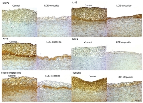
Figure 4 Immunohistochemistry of artery tissues from rabbits treated with saline solution (control group) or LDE-etoposide. Photomicrographs of diaminobenzidine chromogen immunostaining for MMP9, IL-1β, TNF-α, PCNA, topoisomerase IIα, tubulin. Magnifications: 100×.
Abbreviations: IL, interleukin; LDE, cholesterol-rich nanoemulsion; MMP9, matrix metallopeptidase 9; PCNA, proliferating cell nuclear antigen; TNF, tumor necrosis factor.
Abbreviations: CD36, cluster of differentiation 36; LDE, cholesterol-rich nanoemulsion; LDL, low-density lipoprotein; LRP, LDL-related protein; SR-B1, scavenger receptor class B member 1; VSMC, vascular smooth muscle cells.
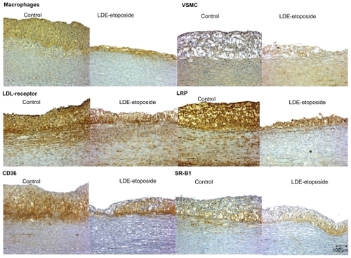
Figure 4 Immunohistochemistry of artery tissues from rabbits treated with saline solution (control group) or LDE-etoposide. Photomicrographs of diaminobenzidine chromogen immunostaining for MMP9, IL-1β, TNF-α, PCNA, topoisomerase IIα, tubulin. Magnifications: 100×.
Abbreviations: IL, interleukin; LDE, cholesterol-rich nanoemulsion; MMP9, matrix metallopeptidase 9; PCNA, proliferating cell nuclear antigen; TNF, tumor necrosis factor.