Figures & data
Figure 1 Gel retardation assay of PEG-g-PEI-SPION/siRNA and scFvCD44v6-PEG-g-PEI-SPION/siRNA polyplexes at various N/P ratios from 2.5 to 20. siRNA bands dissociated from polyplexes were separated by electrophoresis and visualized by an ultraviolet imaging system. Complete siRNA condensation was formed at N/P ratios of 10 and higher. Lane 1, naked siRNA as a control; lanes 2–6, polyplexes formed at N/P ratios of 2.5, 5, 10, 15, and 20.
Abbreviations: PEG, polyethylene glycol; PEI, polyethyleneimine; SPION, superparamagnetic iron oxide nanoparticles; scFvCD44v6, cancer-associated CD44v6 single-chain variable fragment; SiRNA, small interfering RNA.
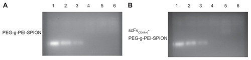
Figure 2 Viability of SGC-7901 cells exposed to PEG-g-PEI-SPION/siRNA and scFvCD44v6-PEG-g-PEI-SPION/siRNA polyplexes for 48 hours at various N/P ratios ranging from 2.5 to 20. Cell viability of targeting and nontargeting polyplexes decreased gradually with increasing N/P ratio. At a higher N/P ratio of 20, cell viability for both groups was still acceptable (over 66%). There was no statistically significant difference between the two groups at individual N/P ratios (P > 0.05). Cell viability is expressed as the mean ± standard deviation of the percentage of absorbance of controls, where 100% equals viability of the control cells. The experiments were carried out in triplicate.
Abbreviations: PEG, polyethylene glycol; PEI, polyethyleneimine; SPION, superparamagnetic iron oxide nanoparticles; scFvCD44v6, cancer-associated CD44v6 single-chain variable fragment; SiRNA, small interfering RNA.
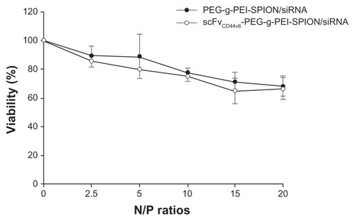
Figure 3 Transfection efficiency and mean fluorescence intensity analyses. Transfection efficiency of Lipofectamine™ (as a control), PEG-g-PEI-SPION, and scFvCD44v6-PEG-g- PEI-SPION at N/P ratios of 5, 10, 15, and 20 were all over 95% (A), while no statistically significant difference between the three siRNA delivery agents was shown for transfection efficiency, but the difference was significant for mean fluorescence intensity (B). Mean fluorescence intensity was highest in the Lipofectamine group, and the mean fluorescence intensity of the scFvCD44v6-PEG-g-PEI-SPION group was higher than in the PEG-g-PEI-SPION group. Especially at N/P ratios of 15 and 20, the difference between the targeting and nontargeting groups was significant (P < 0.05), indicating that the targeting vector transferred more siRNA into cells.
Abbreviations: PEG, polyethylene glycol; PEI, polyethyleneimine; SPION, superparamagnetic iron oxide nanoparticles; scFvCD44v6, cancer-associated CD44v6 single-chain variable fragment.
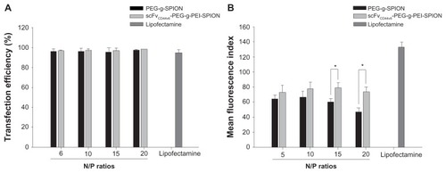
Figure 4 Fluorescence imaging and immunofluorescent staining experiments following siNC-Cy3 transfection. Fluorescence images of SGC-7901 cells transfected with siNCCy3 by scFvCD44v6-PEG-g-PEI-SPION and PEG-g-PEI-SPION at an N/P ratio of 15 were investigated under a fluorescence microscope. As shown in the transfection images, red fluorescence was shown in both groups, but was stronger in the scFvCD44v6-PEG-g-PEI-SPION. After immunofluorescent staining, the red fluorescence of siNC-Cy3 was markedly quenched. Green fluorescence from the fluorescein isothiocyanate-conjugated mouse monoclonal antibody only appeared in the scFvCD44v6-PEG-g-PEI-SPION group, indicating that scFvCD44v6 had successfully attached to PEG-g-PEI-SPION. The merged picture demonstrates that some of the scFvCD44v6-PEG-g-PEI-SPION/siRNA complexes dissociated (red arrows) and some still assembled (white arrows) inside cells.
Abbreviations: PEG, polyethylene glycol; PEI, polyethyleneimine; SPION, superparamagnetic iron oxide nanoparticles; scFvCD44v6, cancer-associated CD44v6 single-chain variable fragment; SiRNA, small interfering RNA.
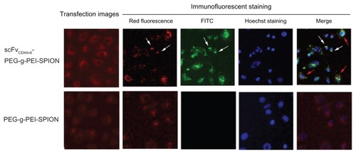
Figure 5 In vivo tumor targeting evaluation. Mice bearing two tumors (SGC-7901 right thigh, A375 left thigh) were used for MRI. Before injection of the SPIO contrast agents, both tumors indicated by red and blue arrows were seen as hyperintense areas in the T2-weighted magnetic resonance images. At 3 hours after injection of scFvCD44v6-PEG-g- PEI-SPION, patchy regions of darkening on the T2-weighted MRI images indicated by green arrows were observed in the SGC-7901 tumor site but not at the A375 tumor site. However, in the PEG-g-PEI-SPION group, T2 signals from the SGC-7901 and A375 tumor sites did not alter before or after injection.
Abbreviations: PEG, polyethylene glycol; PEI, polyethyleneimine; SPION, superparamagnetic iron oxide nanoparticles; scFvCD44v6, cancer-associated CD44v6 single-chain variable fragment; SiRNA, small interfering RNA.
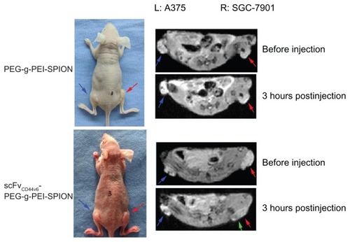
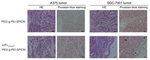
Figure 6 Histology analyses. Hematoxylin and eosin staining and Prussian blue staining were performed on tumor tissues. After Prussian blue staining, blue-positive particles indicated by arrows only appeared in the SGC-7901 tumor area after injection of scFvCD44v6-PEG-g-PEI-SPION, whereas no blue particles were shown at the A375 tumor site in the same mouse. Moreover, in the PEG-g-PEI-SPION group, no SGC-7901 tumor or A375 tumor sites were stained blue following injection.
Abbreviations: PEG, polyethylene glycol; PEI, polyethyleneimine; SPION, superparamagnetic iron oxide nanoparticles; scFvCD44v6, cancer-associated CD44v6 single-chain variable fragment; SiRNA, small interfering RNA.