Figures & data
Figure 1 DSPE-PEG-RGD peptide conjugates (A), Schematic representation of synthesis of DSPE-PEG-RGD peptide conjugates (B) MALDI-TOF mass spectrometric analysis of the DSPE-PEG-RGD conjugate and parental DSPE-PEG maleimide, demonstrating an increase in mass from 2922.79 to 3741.69 after conjugation with the thiolated RGD peptide. This corresponds to one thiolated RGD peptide molecule conjugated to one DSPE-PEG maleimide molecule.
Abbreviations: DSPE-PEG-RGD, Arg(R)-Gly(G)-Asp(D) motif peptide conjugated to 1,2-distearoyl-sn-glycero-3-phosphoethanolamine-N-[maleimide (polyethylene glycol)-2000]; MALDI-TOF, matrix-assisted laser desorption/ionization time-of-flight.
![Figure 1 DSPE-PEG-RGD peptide conjugates (A), Schematic representation of synthesis of DSPE-PEG-RGD peptide conjugates (B) MALDI-TOF mass spectrometric analysis of the DSPE-PEG-RGD conjugate and parental DSPE-PEG maleimide, demonstrating an increase in mass from 2922.79 to 3741.69 after conjugation with the thiolated RGD peptide. This corresponds to one thiolated RGD peptide molecule conjugated to one DSPE-PEG maleimide molecule.Abbreviations: DSPE-PEG-RGD, Arg(R)-Gly(G)-Asp(D) motif peptide conjugated to 1,2-distearoyl-sn-glycero-3-phosphoethanolamine-N-[maleimide (polyethylene glycol)-2000]; MALDI-TOF, matrix-assisted laser desorption/ionization time-of-flight.](/cms/asset/acd701ae-c9cf-49fd-9a5f-fd06df9b0be9/dijn_a_24447_f0001_b.jpg)
Table 1 Particle characterization of siRNA-loaded liposomes
Figure 2 Transmission electron microscopic images of siRNA-loaded liposomes. (A) siRNA-loaded 1 mol% PEGylated liposomes. (B) siRNA-loaded 1 mol% PEGylated, RGD peptide-modified liposomes. The images were taken at 50,000× magnification from the suspended liposomal particles after negative staining. In the bottom-right corner, the local image was enlarged to view the interface of liposomes clearly. Bar = 0.2 μm.
Abbreviations: siRNA, small interfering RNA; PEG, polyethylene glycol.
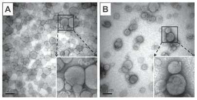
Figure 3 Cell viability of siRNA-loaded liposome preparations in ARPE-19 cells after four hours of treatment: The total lipid concentrations of siRNA-loaded liposome preparations investigated were 51.2, 12.8, and 3.2 μM.
Notes: Data are expressed as the mean ± standard deviation for n = 5. (Student’s t-test *P < 0.05 or **P < 0.01 compared with the untreated group).
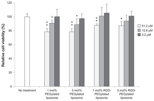
Figure 4 Fluorescence microscopic images showing the internalization of various FAM-siRNA-loaded liposomes in ARPE-19 cells, four hours after administration, at 40× magnification: (A) 1 mol% PEGylated liposomes; (B) 1 mol% PEGylated, RGD peptide-modified liposomes; (C) 5 mol% PEGylated liposomes; (D) 5 mol% PEG ylated, RGD peptide-modified liposomes. SiRNA was labeled with FAM (the arrows are added to point out the green sites), the cell nuclei were stained with Hoechst 33342 (blue), and the cell actin was stained with BODIPY phalloidin (red).
Abbreviations: FAM, fluorescein-labeled; siRNA, small interfering RNA; PEG, polyethylene glycol; RGD, Arg(R)-Gly(G)-Asp(D) motif peptide.
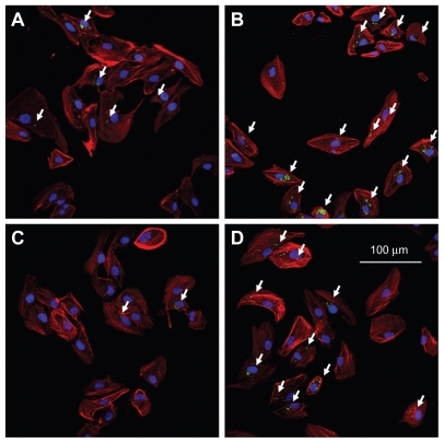
Figure 5 Three-dimensional confocal images showing the intracellular location of FAM-siRNA-loaded liposomes (green) in ARPE-19 cells, four hours after administration, at 100× magnification: 1 mol% PEGylated liposomes (top figure); 1 mol% RGD-PEGylated liposomes (bottom figure). The number of speckled green fluorescence images of FAM-siRNA located within the cytoplasm were counted at each Z-series image (right of figure). The cell nuclei were stained with Hoechst 33342 (blue), and the cell was stained with BODIPY phalloidin (red).
Abbreviations: FAM, fluorescein-labeled; siRNA, small interfering RNA; RGD, Arg(R)-Gly(G)-Asp(D) motif peptide.
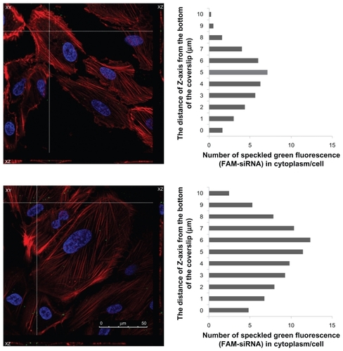
Figure 6 Various amounts of FAM-siRNA loaded liposome uptake as measured by flow cytometry. (A) Forward and side light scatter were used to gate the desired scattered events (normal cells) from dead cells and cell debris. (B) In the normal cells, uptake intensity of FAM-siRNA loaded liposome preparations (green histogram) was compared with the untreated control (block histogram). (C) Fluorescence intensity of FAM-siRNA-loaded liposome preparations in the normal cells expressed as an intensity ratio by dividing by the untreated control.
Notes: Data are expressed as the mean ± SD for N = 3. (Student’s t-test: *P < 0.01 or **P < 0.005, RGD peptide-modified liposomes compared with non-RGD peptide-modified liposomes; 5 mol% compared with 1 mol%).
Abbreviations: FAM, fluorescein-labeled; siRNA, small interfering RNA; SD, standard deviation.
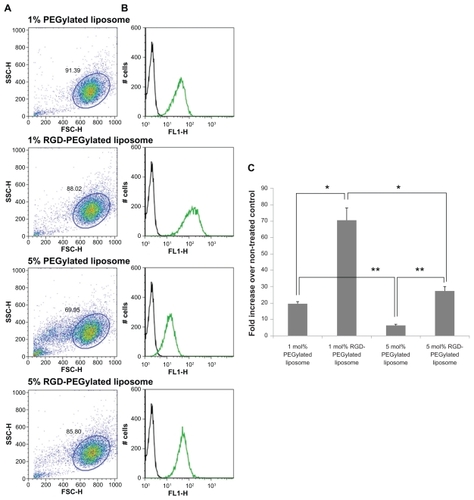
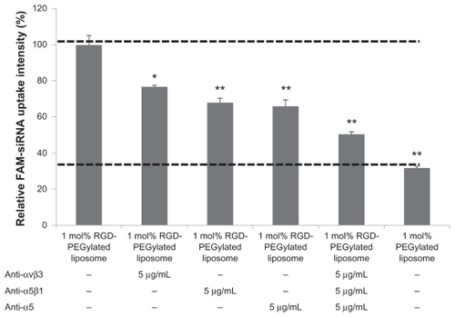
Figure 7 Effect of antibodies on cell integrin receptors and uptake of 1 mol% RGD-PEGylated liposomes by ARPE-19 cells: The cells were incubated with FAM-siRNA -loaded 1 mol% PEGylated liposomes after one hour of blocking with antibody.
Notes: Data are expressed as the mean ± standard deviation for n = 4. (Student’s t-test *P < 0.01, or **P < 0.001, compared with 1 mol% RGD-PEGylated liposomes, no antibody blocking control.)
Abbreviations: FAM, fluorescein-labeled; siRNA, small interfering RNA; RGD, Arg(R)-Gly(G)-Asp(D) motif peptide; PEG, polyethylene glycol.