Figures & data
Figure 1 Illustration for the preparation of camptothecin/iron(III) oxide-embedded poly(D,L-lactide-co-glycolide) composite fibers by electrospinning process.
Abbreviations: CPT, camptothecin; Cu, copper; Fe2O3, iron(III) oxide; PLGA, poly(D,L-lactide-co-glycolide).
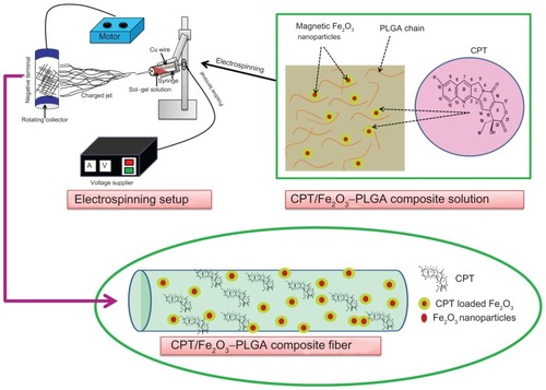
Figure 2 Scanning electron microscopic images of (A and B) pristine poly(D,L-lactide-co-glycolide), (C and D) camptothecin/iron(III) oxide-embedded poly(D,L-lactide-co-glycolide) composite ultrafine fibers, and (E) iron(III) oxide nanoparticles at different magnifications (yellow circles represent point bonding). (F) Energy dispersive X-ray spectrum of camptothecin/iron(III) oxide-embedded poly(D,L-lactide-co-glycolide) composite. The inset photographic images represent the (i) pristine and (ii) composite nano-fibrous mats under ultraviolet fluorescence (λmax 256 nm).
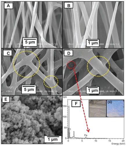
Figure 3 (A) Representative transmission electron microscopic image of the camptothecin/iron(III) oxide-embedded poly(D,L-lactide-co-glycolide) composite. The inset image shows the uniform distribution of iron(III) oxide nanoparticles. Red and blue arrows demonstrate the distinct camptothecin layer and iron(III) oxide nanoparticles, respectively. (B) Electron probe microanalysis mapping result of the camptothecin/iron(III) oxide-embedded poly(D,L-lactide-co-glycolide) composite. The purple square represents the selected area.
Abbreviations: C, carbon; Fe, iron; N, nitrogen; O, oxygen.
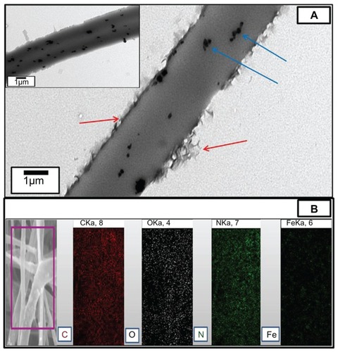
Figure 4 X-ray diffraction patterns of (A) free camptothecin, (B) iron(III) oxide nanoparticles, (C) electrospun pristine poly(D,L-lactide-co-glycolide), and (D) camptothecin/iron(III) oxide-embedded poly(D,L-lactide-co-glycolide) composite fibers.
Note: The red dashed square represents the iron(III) oxide nanoparticle signals.
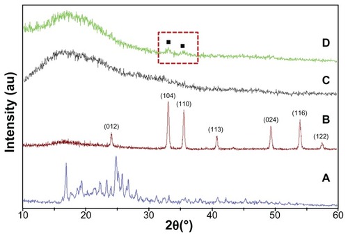
Figure 5 Thermogravimetric analysis graphs of (A) camptothecin/iron(III) oxide-embedded poly(D,L-lactide-co-glycolide) composite and (B) pristine poly(D,L-lactide- co-glycolide) ultrafine fibers.
Note: The inset graph represents the corresponding first derivatives in nitrogen atmosphere.
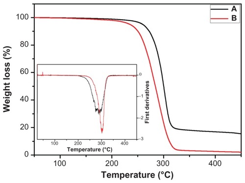
Figure 6 Fourier transform infrared spectra of (A) camptothecin/iron(III) oxideembedded poly(D,L-lactide-co-glycolide) composite and (B) pristine camptothecin/ iron(III) oxide-embedded poly(D,L-lactide-co-glycolide) ultrafine fibers.
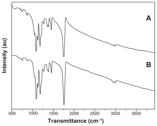
Figure 7 In vitro release profile of (A) camptothecin from electrospun camptothecin/ iron(III) oxide-embedded poly(D,L-lactide-co-glycolide) composite fibers and (B) A355/A368 ratios of camptothecin/iron(III) oxide-embedded poly(D,L-lactide-co- glycolide) composite mat against incubation time in phosphate buffered saline.
Note: The inset scanning electron microscopic image shows the camptothecin/ iron(III) oxide-embedded poly(D,L-lactide-co-glycolide) composite after incubation in phosphate buffered saline at 37°C for 30 days.
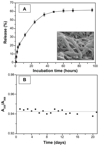
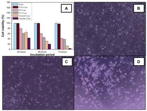
Figure 8 In vitro cytotoxicity of (A) free camptothecin and camptothecin/iron(III) oxide-embedded poly(D,L-lactide-co-glycolide) composite. Untreated C2C12 cells and cells treated with pristine poly(D,L-lactide-co-glycolide) were used as a control. Representative phase contrast images of C2C12 cell lines (B) unexposed to, (C) exposed to 5 μg, and (D) exposed to 10 μg camptothecin/iron(III) oxide-embedded poly(D,L-lactide-co-glycolide) composite.
Note: Magnification 40×.
Abbreviations: CPT, camptothecin; PLGA, poly(D,L-lactide-co-glycolide).