Figures & data
Figure 1 Spectral characteristics of near-infrared-absorbing nanoshells. The absorption spectrum shows the absorbing near-infrared nature (820 nm) of nanoshells with dimensions consisting of a silica core of 100 nm in diameter and shells approximately 10 nm thick. Predicted optical properties were confirmed using ultraviolet-visible spectrophotometry.
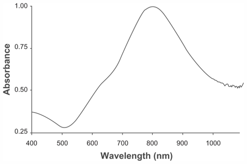
Figure 2 Transmission electron microscopic image of gold-silica nanoshells with an overall diameter of 111 ± 3 nm.
Note: Scale bar = 100 nm.
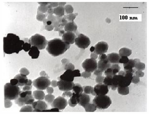
Figure 3 (A) HER2-positive KB cells exposed to anti-HER2 immunonanoshells (nanobody-conjugated nanoshells). (B) Cytotoxicity was observed in cells treated with near-infrared laser. Images represent cells targeted with anti-HER2 nanoshells only.
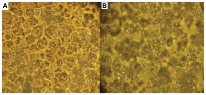
Figure 4 (A) HER2-negative HeLaS3 cells treated with anti-HER2 immunonanoshells. (B) No cytotoxicity was observed in HeLaS3 cells following near-infrared laser treatment.
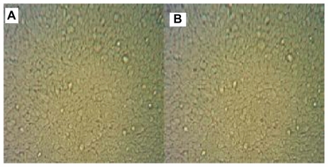
Figure 5 MTT assay results for normal HER2-positive (KB) and HER2-negative (HeLaS3) cells before and after near-infrared laser irradiation.
Abbreviation: NIR, near-infrared.
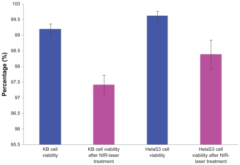
Figure 6 MTT assay results for KB and HeLaS3 cells following treatment with HER2-targeted nanobodies and near-infrared laser.
Abbreviation: NIR, near-infrared.
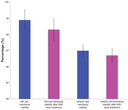
Figure 7 MTT assay results for KB and HeLaS3 cells exposed to bare nanoshells. Both cell types were treated with a near-infrared laser.
Abbreviation: NIR, near-infrared.
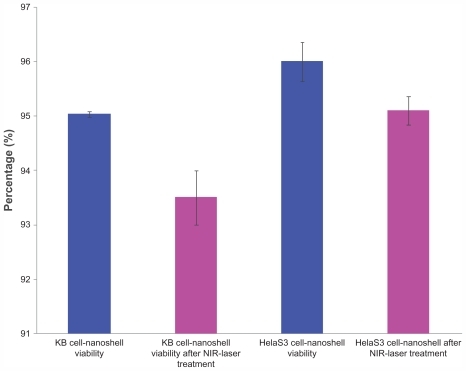
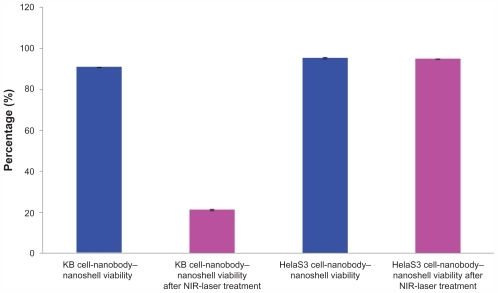
Figure 8 Anti-HER2 immunonanoshells (nanobody-conjugated nanoshells) were added to KB and HeLaS3 cells, and the cells were treated with an near-infrared laser. An MTT assay was performed on both cell types and our results indicated that immunotargeted nanoshells can selectively induce specific cell death in vitro.
Abbreviation: NIR, near-infrared.