Figures & data
Figure 1 (A) Scanning electron microscopy image of POH-loaded PLGA microparticles. (B) Corresponding particle analyzer data as obtained by Nanophox (Sympatec GmbH, Clausthal-Zellerfeld, Germany) particle size analyzer. (C) In vitro release kinetics from PLGA microparticles. Release kinetics of POH-PLGA microparticle formulation demonstrates ∼30% release of POH from the copolymer at 168 hours, when formulation was dispersed in 20 mM phosphate buffer, pH 7.4 at 37°C.
Note: Data are means ± standard deviations of three independent experimental values.
Abbreviations: PLGA, poly-lactic glycolic acid; POH, perillyl alcohol; EHT, extra high tension; WD, working distance.
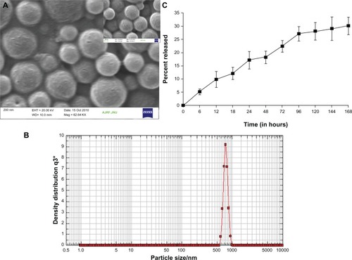
Figure 2 Erythrocyte lysis test: In vitro toxicity was measured by erythrocyte lysis caused by different POH formulations.
Notes: Hemolysis test was performed as described in the Materials and methods section. Data represented here are means of three different experiments ± standard deviations.
Abbreviations: DMSO, dimethyl sulfoxide; PLGA, poly-lactic glycolic acid; POH, perillyl alcohol.
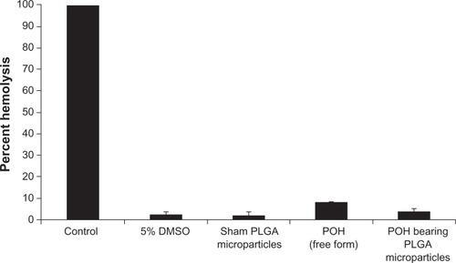
Table 1 Concentrations of creatinine and ALP in plasma of animals treated with POH-bearing PLGA microparticle formulation
Figure 3 In vitro cytotoxicity of perillyl alcohol against A253 cell line as revealed by MTT assay.
Notes: MTT assay was performed as described in the Materials and methods section. Data represented here are means of three different experiments ± standard deviations.
Abbreviation: MTT, 3-(4,5-dimethylthiazol-2-yl)-2,5-diphenyltetrazolium bromide.
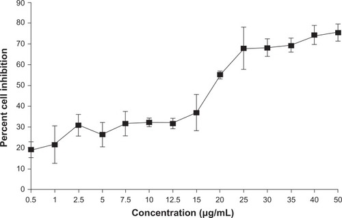
Figure 4 Determination of differential cytotoxicity of various formulations of POH using MTT assay. Cells were incubated with various formulations of POH for 48 hours.
Notes: The percentage cell viability was measured with MTT assay as described in the Materials and methods section. Data represented here are means of three different experiments ± standard deviations (POH-PLGA microparticle versus free form POH P < 0.001).
Abbreviations: MTT, 3-(4,5-dimethylthiazol-2-yl)-2,5-diphenyltetrazolium bromide; PLGA, poly-lactic glycolic acid; POH, perillyl alcohol.
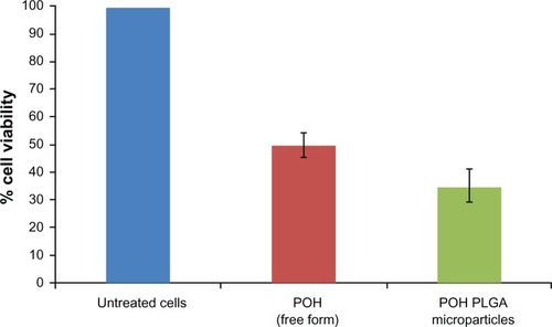
Figure 5 Effect of POH-bearing formulations on expression of pro-apoptotic molecules. A253 cancer cells were treated with POH-bearing microformulations for different time periods, and cell lysates were used to examine the expression of apoptotic molecules. (A) Expression profile of apoptotic factors at 12 hours post-incubation. (B) Expression profile of apoptotic factors at 24 hours post-incubation.
Abbreviations: PLGA, poly-lactic glycolic acid; POH, perillyl alcohol.
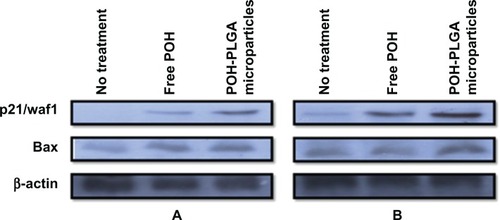
Figure 6 Chemotherapeutic effect of two different formulations of POH in regression of tumors in treated animals.
Notes: Percentage regression was calculated to analyze the most effective formulation of POH. It was assessed by measuring the size with Vernier calipers, and tumor volume was calculated as given in the Materials and methods section (POH-PLGA microparticles versus free form POH; P < 0.001). Sham-PLGA microparticles behaved as control.
Abbreviations: PLGA, poly-lactic glycolic acid; POH, perillyl alcohol.
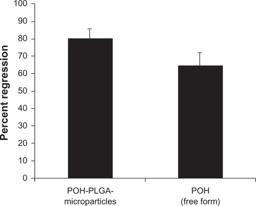
Figure 7 Effect of POH-bearing PLGA formulations on survival of DMBA-induced skin papilloma-carrying animals.
Notes: Kaplan-Meier graph shows the efficacy of various POH formulations in terms of percentage survival after treatment at different time intervals. The study was continued for a period of 13 weeks as described in the Materials and methods section (POH-PLGA microparticles versus free form POH; P < 0.05).
Abbreviations: PLGA, poly-lactic glycolic acid; POH, perillyl alcohol.
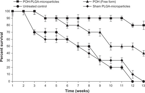
Figure 8 Photomicrographs of hematoxylin/eosin-stained sections, showing: (A) normal smooth skin of healthy animals with keratinocytes of only a few cell layers thick, mild keratin, and pilosebaceous units; (B) the positive control (DMBA treated animals followed by no POH treatment) has profuse papillomatous growth with complex fibrovascular core, prominent acanthosis, and keratin perl; (C) treatment with free form of POH resulted in large amount of keratin, and marked acanthosis; while (D) the treatment with PLGA microsphere encapsulated POH resulted in hyperkeratosis, mild acanthosis, and thin but long papillary growth.
Note: Magnification 100×.
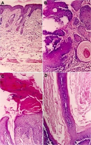
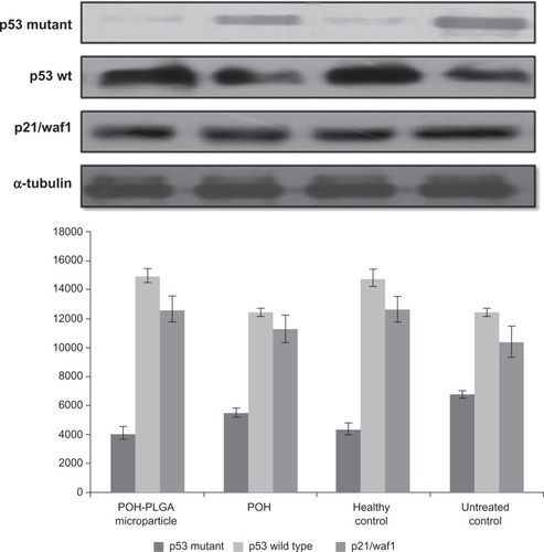
Figure 9 Effect of different POH-bearing formulations on the expression of different apoptosis-regulating molecules in treated animals.
Notes: Skin papilloma cell lysates were prepared as described in the Materials and methods section. Cell lysate was resolved and analyzed using SDS-PAGE. To ensure equal loading, membranes were re-probed for the presence of tubulin, a housekeeping protein, using α-tubulin antibody.
Abbreviations: PLGA, poly-lactic glycolic acid; POH, perillyl alcohol.