Figures & data
Table 1 Physical properties of pre- and post-functionalized SWCNTs
Figure 1 Transmission electron microscopy of SWCNTs pre- and post-functionalization. SWCNTs before (A) and after (B) oxidization using HNO3:H2SO4. Treatment with acid resulted in chemical modifications of the SWCNTs and the formation of carboxyl groups on the surface. Inset: tube containing SWCNTs, showing the enhancement of dispersibility of SWCNTs 2 weeks post-functionalization.
Abbreviation: SWCNT, single-walled carbon nanotube.
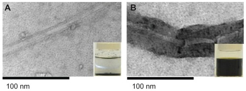
Figure 2 HPA-lectin-FITC-SWCNTs. A sample of functionalized SWCNTs conjugated to HPA-FITC was viewed under the confocal microscope, in transmission mode (A) and in fluorescence mode, following excitation at 488 nm (B). The results show that a proportion of the SWCNTs had been successfully conjugated to the FITC-labeled lectin.
Abbreviations: FITC, fluorescein isothiocyanate; HPA, Helix pomatia agglutinin; SWCNT, single-walled carbon nanotube.
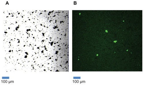
Figure 3 The effect of different acid exposure times on attachment of lectin to SWCNTs. The SWCNTs were treated with acid for 0.5, 1, 2, 3, 4, and 5 hours, followed by HPA-lectin-FITC conjugation. Confocal microscopy was used to determine the relative fluorescence for the SWCNTs prepared. An increase in the fluorescence was observed as the exposure time of SWCNTs to HNO3 and H2SO4 increased. The relative fluorescence following 0.5 hours treatment with HNO3 and H2SO4 was compared with longer incubation periods.
Notes: Statistical analysis was performed using the Student’s t-test, and the P-values are indicated. Values are mean ± standard deviation. *P < 0.05 (2 hrs); **P < 0.01 (3 hrs, 4 hrs and 5 hrs).
Abbreviations: FITC, fluorescein isothiocyanate; HPA, Helix pomatia agglutinin; SWCNT, single-walled carbon nanotube.
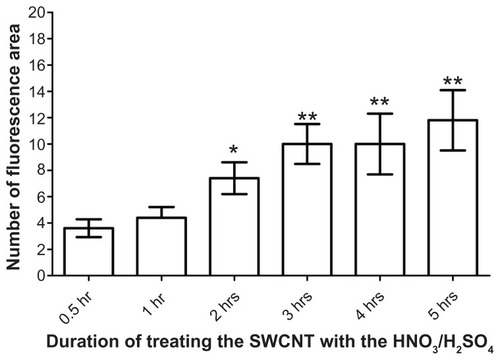
Figure 4 Fourier transform infrared spectroscopy analysis of SWCNTs. Before (red) and after (blue) functionalization. Following functionalization, peaks observed at 1750 cm−1 and 3450 cm−1 indicate that −COOH groups had been successfully grafted onto the surface of the SWCNTs.
Abbreviation: SWCNT, single-walled carbon nanotube.
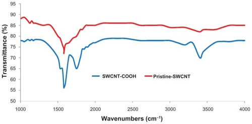
Figure 5 Confocal microscopy of MCF-7 breast cancer cells incubated with HPA-FITC and HPA-lectin-FITC linked to SWCNTs. Lectin binding to MCF-7 and SW480 cells as viewed using a Leica TCS SP2 confocal microscope. Cells were incubated with FITC-labeled HPA (green) and lectin-SWCNTs (green), and the nuclei were counterstained with To-Pro®3 (blue). MCF-7 were observed to interact with the HPA lectin either linked to FITC or to SWCNT; in contrast, the SW480 cells did not bind the lectin.
Abbreviations: FITC, fluorescein isothiocyanate; HPA, Helix pomatia agglutinin; SWCNT, single-walled carbon nanotube.
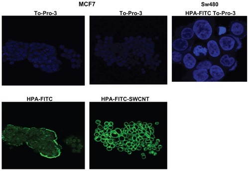
Figure 6 Temperature profile of SWCNTs, pre- and post-functionalization. Following exposure to light at λ 808 nm, functionalized SWCNTs achieved a higher temperature profile compared with non-functionalized CNTs.
Note: Mean values ± standard deviation are shown.
Abbreviations: CNT, carbon nanotube; SWCNT, single-walled CNT; F-SWCNT, functionalized SWCNT.
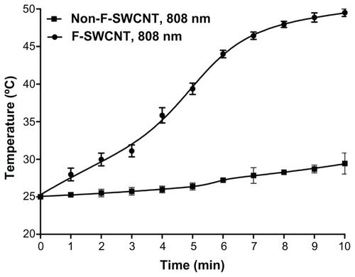
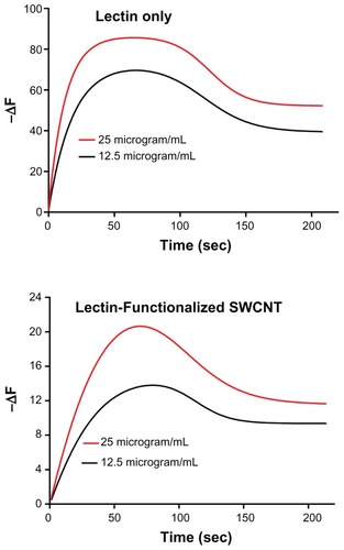
Figure 7 Sensorgrams illustrating the binding profile of HPA-FITC and HPA-lectin-SWCNTs to MCF-7 cells grown on a biosensor chip surface. The association (85 second) and dissociation (165 second) phases were monitored giving a change in the resonance frequency (ΔF) as the lectin interacted with the surface of the MCF-7 cells on the chip surface. The lectin interaction with MCF-7 cells was investigated at 12.5 μg/mL and 25 μg/mL as shown, using HPA alone and HPA-lectin-SWCNTs. The flow rate was maintained at 25 μL/min, and the temperature was 22°C. The binding profile for native HPA was comparable to the profile obtained for HPA-lectin-SWCNTs, indicating that the conjugation process did not affect the rate of epitope binding by the lectin.
Abbreviations: FITC, fluorescein isothiocyanate; HPA, Helix pomatia agglutinin; SWCNT, single-walled carbon nanotube.