Figures & data
Figure 1 Structure of paclitaxel and PGG-paclitaxel conjugate.
Abbreviations: PTX, paclitaxel; PGG, poly(L-γ-glutamylglutamine).
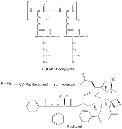
Figure 2 Thermogravimetric/differential thermal analyzer curve of free paclitaxel (A) and polymer PGG-PTX conjugate (B) with weight percent plotted against temperature.
Abbreviations: PTX, paclitaxel; PGG, poly(l-γ-glutamylglutamine); TGA, thermogravimetric analysis; DTA, differential thermal analysis.
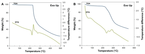
Figure 3 Transmission electron micrograph of poly(l-γ-glutamylglutamine)-paclitaxel nanoconjugates.
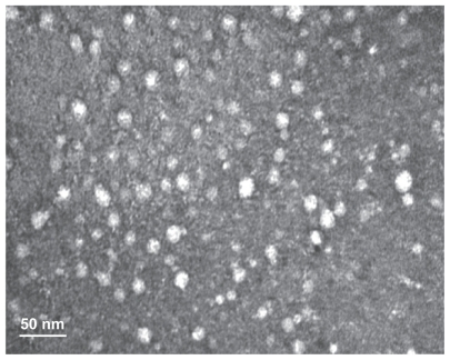
Figure 4 Stability of poly(l-γ-glutamylglutamine)-paclitaxel as a function of (A) paclitaxel release and (B) particle size at room temperature and 4°C over a period of three months.
Note: Each point represents the mean of three samples with the standard error of the mean (vertical bars).
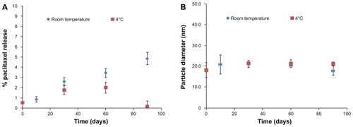
Figure 5 Fluorescence microscopy images showing internalization of NCI-H460 cell treated with coumarin 6-labeled poly(l-γ-glutamylglutamine)-paclitaxel nanoparticles (1 mg/mL) at 37°C for 30 minutes and 4 hours. On the left, the green-labeled corresponds to fluorescent poly(l-γ-glutamylglutamine)-paclitaxel loaded with coumarin 6. On the right, merged images of the nuclei stained with DAPI (blue) and of the cells uptake of coumarin 6-loaded poly(l-γ-glutamylglutamine)-paclitaxel (green).
Notes: The results are representative of three independent experiments. Magnification 200×.
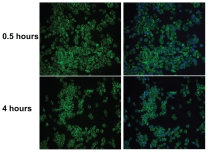
Figure 6 Cellular uptake of coumarin 6-loaded poly(l-γ-glutamylglutamine)-paclitaxel nanoparticles at (A) different concentrations incubated at 37°C for 4 hours, and (B) for different incubation periods of time at 5 mg poly(l-γ-glutamylglutamine)-paclitaxel per mL. Cell-associated fluorescence was measured at the excitation and emission wavelengths of 430 and 485 nm, respectively.
Note: Results are expressed as the mean of three independent experiments ± standard deviation (n = 3–4).
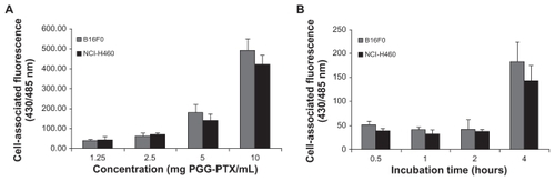
Table 1 IC50 (μg/mL) of free paclitaxel and PGG-paclitaxel in various cell lines
Figure 7 Cells were seeded in 96-well cell culture plates overnight before exposure to poly(l-γ-glutamylglutamine)-paclitaxel at a concentration range of 0.01–10 mg/mL. Incubation continued for (A) 48 hours and (B) 72 hours before MTS staining. Survival of treated cells compared with untreated controls at each time point is shown.
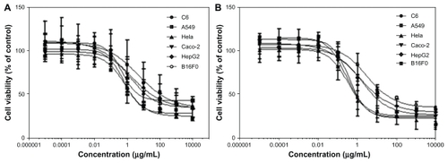
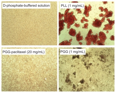
Figure 8 Effects of poly(L-γ-glutamylglutamine)-paclitaxel conjugate and poly(L-γ-glutamylglutamine) on red blood cell aggregation in pH 7.4 phosphate-buffered solution.
Figure 9 Effects of PGG-paclitaxel, PGG, and PLL on red blood cell aggregation in pH 7.4 phosphate-buffered solution.
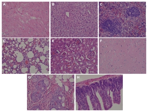
Figure 10 H460 cells were injected into each shoulder of 6–8-week-old female athymic nude (nu/nu) mice. These mice were treated with a single intravenous injection of PGG-PTX (350 mg/mL, paclitaxel equivalent) when tumor size reached an average volume of 100 mm3. Two mice were sacrificed after treatment, and sections of various tissues were examined by using hematoxylin and eosin staining. (A) heart, (B) liver, (C) spleen, (D) lung, (E) kidney, (F) brain, (G) skin, and (H) small intestine.
Note: Magnification 100×.
Abbreviations: PGG, poly(L-γ-glutamylglutamine); PTX, paclitaxel.
Note: The results are representative of three independent experiments.
Abbreviations: PGG, poly(L-γ-glutamylglutamine); PLL, poly(L-lysine).
Note: The relative percentages of hemolysis are shown as the mean ± standard deviation (n = 3).
Abbreviations: PBS, phosphate-buffered solution; PGG, poly(L-γ-glutamylglutamine).
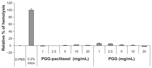
Figure 9 Effects of PGG-paclitaxel, PGG, and PLL on red blood cell aggregation in pH 7.4 phosphate-buffered solution.
Figure 10 H460 cells were injected into each shoulder of 6–8-week-old female athymic nude (nu/nu) mice. These mice were treated with a single intravenous injection of PGG-PTX (350 mg/mL, paclitaxel equivalent) when tumor size reached an average volume of 100 mm3. Two mice were sacrificed after treatment, and sections of various tissues were examined by using hematoxylin and eosin staining. (A) heart, (B) liver, (C) spleen, (D) lung, (E) kidney, (F) brain, (G) skin, and (H) small intestine.
Note: Magnification 100×.
Abbreviations: PGG, poly(L-γ-glutamylglutamine); PTX, paclitaxel.
Note: The results are representative of three independent experiments.
Abbreviations: PGG, poly(L-γ-glutamylglutamine); PLL, poly(L-lysine).
Figure 10 H460 cells were injected into each shoulder of 6–8-week-old female athymic nude (nu/nu) mice. These mice were treated with a single intravenous injection of PGG-PTX (350 mg/mL, paclitaxel equivalent) when tumor size reached an average volume of 100 mm3. Two mice were sacrificed after treatment, and sections of various tissues were examined by using hematoxylin and eosin staining. (A) heart, (B) liver, (C) spleen, (D) lung, (E) kidney, (F) brain, (G) skin, and (H) small intestine.
Note: Magnification 100×.
Abbreviations: PGG, poly(L-γ-glutamylglutamine); PTX, paclitaxel.