Figures & data
Figure 1 Schematic diagram of the synthesis of PCL-Tween 80 copolymer.
Abbreviation: PCL, poly-ɛ-caprolactone.
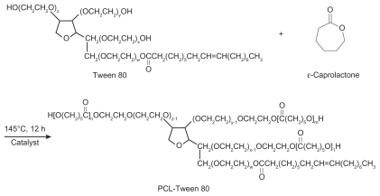
Figure 2 Typical 1H-NMR spectra of PCL-Tween 80 copolymer.
Abbreviations: PCL, poly-ɛ-caprolactone; NMR, nuclear magnetic resonance.
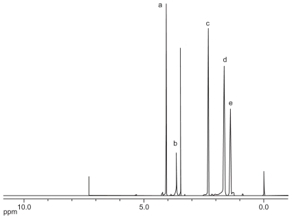
Figure 3 Fourier transform infrared spectra of PCL-Tween 80 copolymer.
Abbreviation: PCL, poly-ɛ-caprolactone.
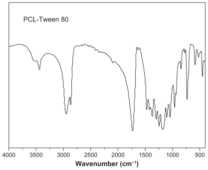
Figure 4 Thermogravimetric profiles of PCL-Tween 80 copolymer.
Abbreviation: PCL, poly-ɛ-caprolactone.
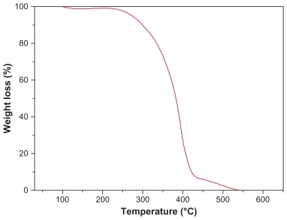
Table 1 Characterization of nanoparticles (n = 3)
Figure 5 Field emission scanning electron microscopy image of docetaxel-loaded PCL-Tween 80 nanoparticles.
Abbreviation: PCL, poly-ɛ-caprolactone.
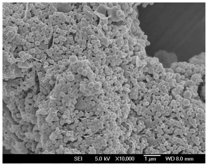
Figure 6 In vitro release profile of docetaxel-loaded PCL-Tween 80 nanoparticles and docetaxel-loaded PCL nanoparticles.
Abbreviations: PCL, poly-ɛ-caprolactone; NPs, nanoparticles.
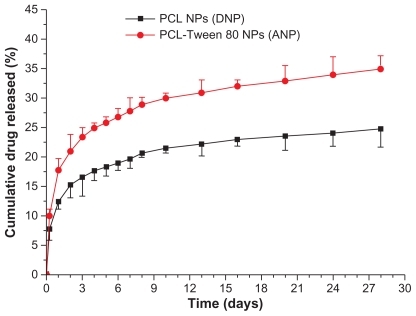
Figure 7 Cellular uptake of coumarin 6-loaded PCL-Tween 80 nanoparticles and coumarin 6-loaded PCL nanoparticles by C6 cells after 2 hours of incubation.
Note: *P < 0.05.
Abbreviation: PCL, poly-ɛ-caprolactone.
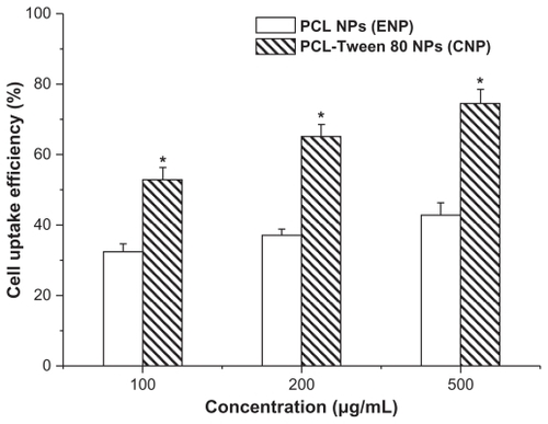
Figure 8 Confocal laser scanning microscopy images of C6 cells after 2 hours of incubation with coumarin 6-loaded PCL-Tween 80 nanoparticles at 37.0°C. The cells were stained by DAPI (blue) and the coumarin 6-loaded nanoparticles are green. The cellular uptake was visualized by overlaying images obtained by green fluorescent protein filter and DAPI filter: left image from DAPI channel (A); center image from green fluorescent protein channel (B); right image from combined green fluorescent protein channel and DAPI channel (C).
Abbreviation: PCL, poly-ɛ-caprolactone.
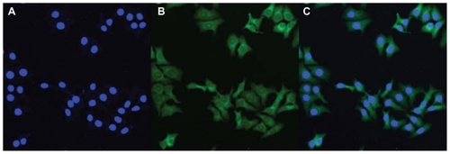
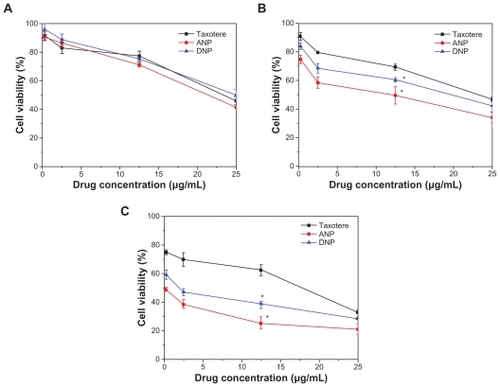
Figure 9 Viability of C6 cells after 24 (A), 48 (B), and 72 (C) hours of cell culture with docetaxel formulated in ANP and DNP in comparison with Taxotere® at the same docetaxel dose.
Note: n = 6, *P < 0.05.
Abbreviations: PCL, poly-ɛ-caprolactone; ANP, PCL-Tween 80 nanoparticles; DNP, PCL nanoparticles.