Figures & data
Table 1 Properties of SPIONs with different formulations
Figure 1 Synthetic approach of folate-PEG-PCL for SPION coating.
Abbreviations: PEG-PCL, poly(ethylene glycol)-poly(ɛ-caprolactone); SPION, superparamagnetic iron oxide nanoparticle.
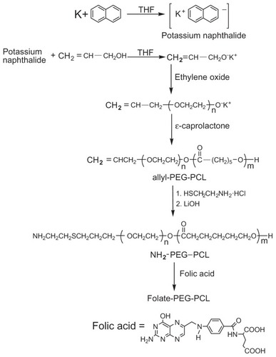
Figure 2 Synthesis of monodisperse Fe3O4 nanoparticles coated with biodegradable diblock copolymer.
Abbreviation: PEG-PCL, poly(ethylene glycol)-poly(ɛ-caprolactone).
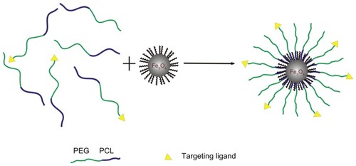
Figure 3 Transmission electron microscopy (TEM) images of synthesized 6 nm SPIONs (A) and PEG-PCL-SPIONs (B). The insert (a) shows the synthesized SPIONs were soluble in hexane, and PEG-PCL-SPIONs were easily dispersed in water (b). The selected area electron diffraction (SAED) pattern of 6-nm Fe3O4 nanoparticles is shown in (C).
Abbreviation: PEG-PCL-SPIONs, poly(ethylene glycol)-poly(ɛ-caprolactone) superparamagnetic iron oxide nanoparticles.
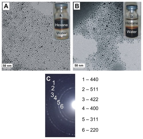
Figure 4 DLS profiles of PEG-PCL-SPIONs and blank PEG-PCL micelle.
Abbreviations: DLS, dynamic light scattering; PEG-PCL-SPIONs, poly(ethylene glycol)-poly(ɛ-caprolactone) superparamagnetic iron oxide nanoparticles.
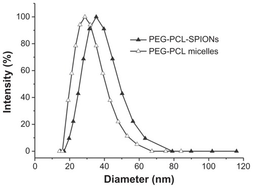
Figure 5 Hysteresis loops of 6-nm Fe3O4 nanoparticles (A) and PEG-PCL-SPIONs (B) measured at 300 K and 10 K.
Abbreviation: PEG-PCL-SPIONs, poly(ethylene glycol)-poly(ɛ-caprolactone) superparamagnetic iron oxide nanoparticles.
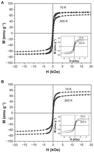
Table 2 Dependence of MRI T2-weighted signal intensity and signal intensity changes of tumor on the postinjection time
Figure 6 T2-weighted MRI images (TR/TE, 5000/100 ms) taken at 0 and 3 hours after injecting 5 mg of Fe/kg of PEG-PCL-SPIONs (A and B) and Fa-PEG-PCL-SPIONs (C and D) via tail vein into a nude mouse bearing BEL-7402 tumor (about 1 cm in diameter).
Note: The white arrow indicates the xenograft tumor for determining T2-weighted signal intensity change.
Abbreviations: MRI, magnetic resonance imaging; TR, repetition time; TE, echo time; Fa-PEG-PCL-SPIONs, folate-attached poly(ethylene glycol)-poly(ɛ-caprolactone) superparamagnetic iron oxide nanoparticles.
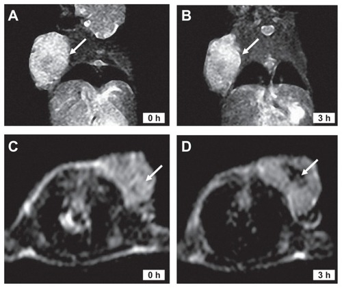
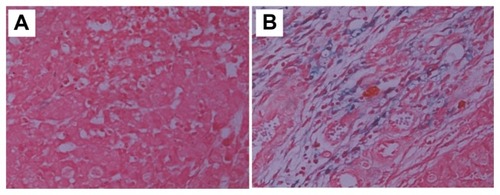
Figure 7 Prussian blue staining images of tumor tissues taken from mice at a time point 3 hours after injection of PEG-PCL-SPIONs (A) and Fa-PEG-PCL-SPIONs (B).
Note: Blue stain density reflects the level of SPIO accumulation within tumor.
Abbreviation: Fa-PEG-PCL-SPIONs, folate-attached poly(ethylene glycol)-poly(ɛ-caprolactone) superparamagnetic iron oxide nanoparticles.