Figures & data
Table 1 Surface modification and zeta potential of PEGylated QD (composed of CdSe/ZnS with core size of 5.3 nm and hydrodynamic size of 25 nm)
Figure 1 Fluxes of amine-, carboxylate-, or non-modified quantum dots across rat alveolar epithelial cell monolayers over 24 hours are not significantly different from each other.
Note: Apical [quantum dot] = 6.25 μg/mL (n = 9).
![Figure 1 Fluxes of amine-, carboxylate-, or non-modified quantum dots across rat alveolar epithelial cell monolayers over 24 hours are not significantly different from each other.Note: Apical [quantum dot] = 6.25 μg/mL (n = 9).](/cms/asset/02cfec0b-96ef-403b-943a-8e9531f5f305/dijn_a_26051_f0001_b.jpg)
Figure 2 Fluxes of quantum dots across rat alveolar epithelial cell monolayers treated with 2 mM EGTA for 24 hours increased significantly (by about 50%–130%) from respective controls. Transmonolayer resistance of rat alveolar epithelial cell monolayers decreased by about 95% during experiments in the presence of EGTA.
Notes: Apical [quantum] = 6.25 μg/mL. *Significantly increased compared with control (n = 6).
![Figure 2 Fluxes of quantum dots across rat alveolar epithelial cell monolayers treated with 2 mM EGTA for 24 hours increased significantly (by about 50%–130%) from respective controls. Transmonolayer resistance of rat alveolar epithelial cell monolayers decreased by about 95% during experiments in the presence of EGTA.Notes: Apical [quantum] = 6.25 μg/mL. *Significantly increased compared with control (n = 6).](/cms/asset/32485b0e-624d-4fc6-a31b-beabcd340557/dijn_a_26051_f0002_b.jpg)
Figure 3 Fluxes of quantum dots across rat alveolar epithelial cell monolayers increased significantly (by about 85%) when temperature decreased from 37°C to 4°C. Transmonolayer resistance of rat alveolar epithelial cell monolayers decreased by about 95% at 4°C.
Notes: Apical [quantum dot] = 6.25 μg/mL. *Significantly increased from control
![Figure 3 Fluxes of quantum dots across rat alveolar epithelial cell monolayers increased significantly (by about 85%) when temperature decreased from 37°C to 4°C. Transmonolayer resistance of rat alveolar epithelial cell monolayers decreased by about 95% at 4°C.Notes: Apical [quantum dot] = 6.25 μg/mL. *Significantly increased from control](/cms/asset/d6d4ac2c-e776-40c4-a719-77d96aa5f991/dijn_a_26051_f0003_b.jpg)
Figure 4 Fluxes of quantum dots (6.25 μg/mL apical concentration) did not decrease significantly when rat alveolar epithelial cell monolayers were treated with 200 μm methyl-β-cyclodextrin for 24 hours. Transmonolayer resistance was not significantly affected by the presence of methyl-β-cyclodextrin compared with control (n = 6).
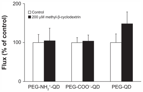
Figure 5 Fluxes of quantum dots (6.25 μg/mL apical concentration) did not decrease in the presence of 28 μM chlorpromazine for 24 hours. Transmonolayer resistance of rat alveolar epithelial cell monolayers after 24 hours of exposure to chlorpromazine was not different from control (n = 6).
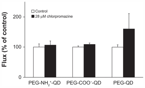
Figure 6 Fluxes of quantum dots (6.25 μg/mL apical concentration) did not decrease in the presence of 80 μM dynasore for 24 hours. Transmonolayer resistance of rat alveolar epithelial cell monolayers was increased by about 30% after 24 hours of exposure to dynasore compared with control.
Note: *Significantly increased from control (n = 6).
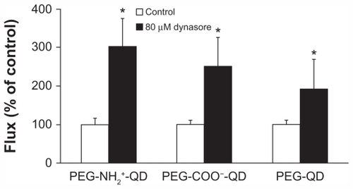
Figure 7 Confocal photomicrograph showing amine-modified PEGylated quantum dots (red) internalized into alveolar epithelial cells following 24 hours of apical exposure to 6.25 μg/mL quantum dots. Zonula occludens-1 is shown in green and nuclei are shown in blue.
