Figures & data
Figure 1 Skin microstructure and skin penetration mechanisms of nanoparticles. (A) Penetration trough sebaceous glands and hair follicles. (B) Transcellular penetration. (C) Intracellular penetration.
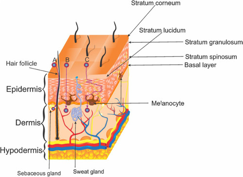
Figure 2 Melanoma stages.
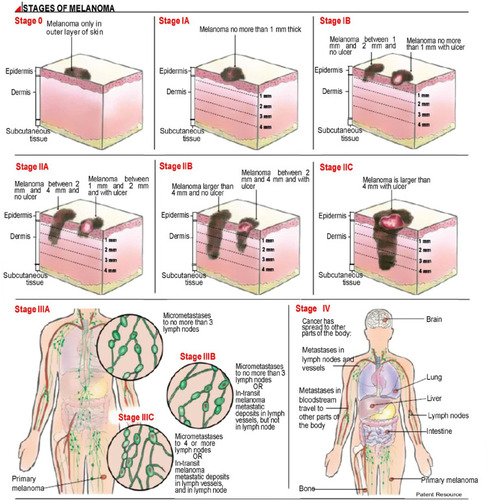
Figure 3 (A) Schematic representation of a liposome. (B) Classification by vesicle number.
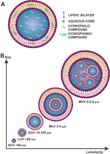
Figure 4 Main components of liposomes. (A) Phospholipids (nomenclature=phosphatidyl + choline, + ethanolamine, + glycerol, + inositol, + serine) the molecular weight of the lipids will change depending on the R groups; (B) Chemical structure of cholesterol.
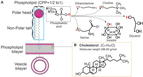
Table 1 Physical Properties of Liposomes with Different Compounds and Lipid Ratio After Being Prepared with Thin-Film Hydration
Figure 5 Fluorescent images of Nile red stained giant unilamellar liposomes obtained by confocal laser scanning microscopy (multiphoton confocal microscope LSM 710 NLO, Carl Zeiss.
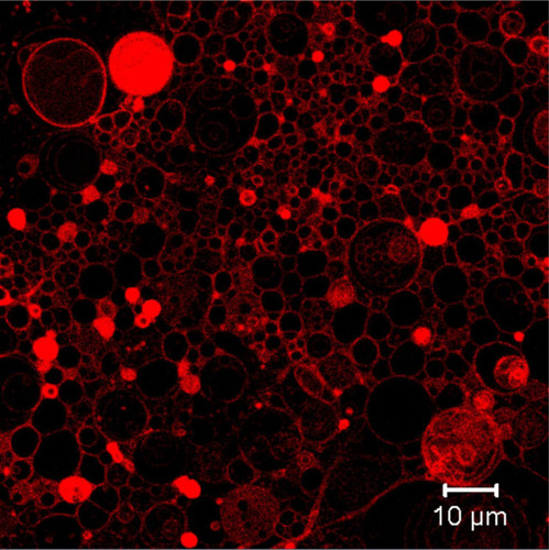
Table 2 Liposomal Formulations for Skin Delivery of Different Compounds
Figure 6 Chemical structures of bioactive compounds that have been incorporated into liposomes.
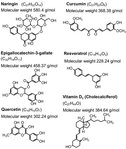
Figure 7 Chemical structures of most used edge activators in the preparation of liposomes.
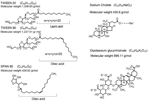
Table 3 Patents for Liposome Formulation in Skincare
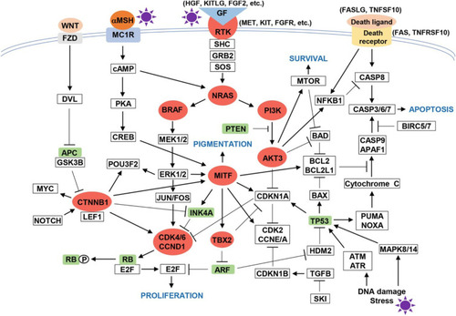
Figure 8 Melanomagenesis pathway.