Figures & data
Table 1 Schematic description of size and surface characteristics of SPIONs tested
Figure 1 Physicochemical characterization of SPIONs tested. TEM images of bare SPIONs (A) and SPIONs modified with TEOS (B), APTMS (C), TEOS-APTMS (D), or citrate (E), along with the zeta-potentials (F) of each SPION.
Note: The TEM images shown in this figure are representative of six independent experiments with similar results.
Abbreviations: APTMS, (3-aminopropyl)trimethoxysilane; SPION, superparamagnetic iron oxide nanoparticles; TEM, transmission electron microscopy; TEOS, tetraethyl orthosilicate.
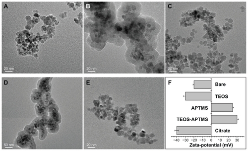
Figure 2 Effect of SPIONs on mitochondrial activity. Relative cell viability of L-929 cells exposed for 24 hours to increasing concentrations (0~1000 ppm) of SPIONs coated with various functional groups was evaluated using the WST-8 assay.
Abbreviations: APTMS, (3-aminopropyl)trimethoxysilane; SPION, superparamagnetic iron oxide nanoparticles; TEOS, tetraethyl orthosilicate.
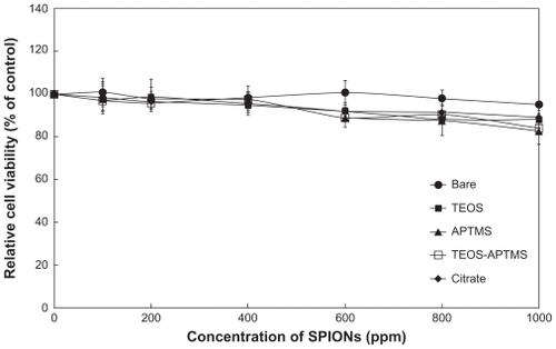
Figure 3 Effects of SPIONs on oxidative stress and cell membrane integrity. Intracellular ROS levels (A) and LDH release profiles (B) in L-929 cells exposed to increasing concentrations (0~1000 ppm) of SPIONs coated with various functional groups for 24 hours were evaluated by the DCF and LDH assays, respectively.
Abbreviations: APTMS, (3-aminopropyl)trimethoxysilane; DCF, 2′,7′-dichlorodihydrofluorescein; LDH, lactate dehydrogenase; ROS, reactive oxygen species; SPION, superparamagnetic iron oxide nanoparticles; TEOS, tetraethyl orthosilicate.
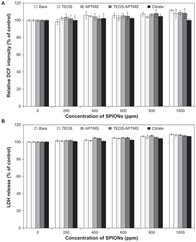
Figure 4 Effects of SPIONs on DNA stability. Tail content of DNA in L-929 cells exposed to increasing concentrations (0–1000 ppm) of SPIONs coated with various functional groups for 24 hours was determined by the comet assay.
Notes: *P < 0.05 vs nontreated control; #P < 0.05 vs SPION-treated cells with 100 ppm.
Abbreviations: APTMS, (3-aminopropyl)trimethoxysilane; DMSO, dimethyl sulfoxide; SPION, superparamagnetic iron oxide nanoparticles; TEOS, tetraethyl orthosilicate.
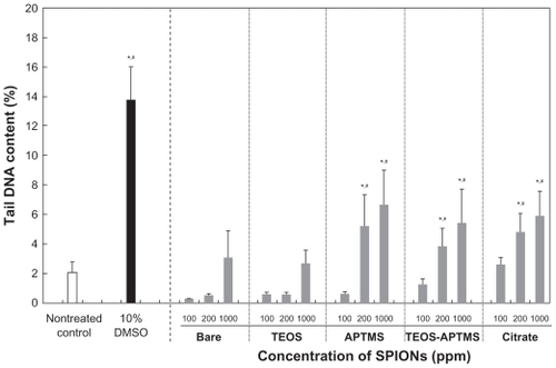
Figure 5 Electron microscopy images of L-929 cells. Morphological alterations and intracellular ultrastructures of L-929 cells exposed to 500 ppm of SPIONs coated with various functional groups for 24 hours were observed by SEM (A) and TEM (B), respectively.
Notes: The scale bars in the large and inserted SEM images were 30 μm and 6 μm, respectively. The scale bars in the large and inserted TEM images were 5000 nm and 1000 nm, respectively. The electron micrographs shown in this figure are representative of six independent experiments with similar results.
Abbreviations: APTMS, (3-aminopropyl)trimethoxysilane; DMSO, dimethyl sulfoxide; SEM, scanning electron microscopy; SPION, superparamagnetic iron oxide nanoparticles; TEM, transmission electron microscopy; TEOS, tetraethyl orthosilicate.
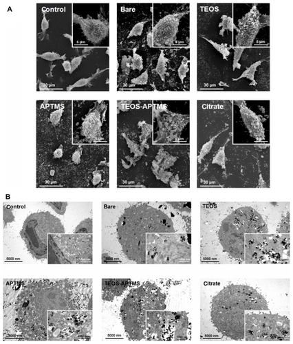
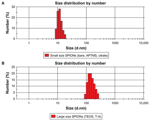
Figure S1 Size distributions of relatively small SPIONs (ie, bare and APTMS- or citrate-coated) (A) and large SPIONs (ie, TEOS- or T-A-coated) (B) in aqueous solution.
Note: The Zetasizer histograms shown in this figure are representative of five independent experiments with almost similar results.
Abbreviations: APTMS, (3-aminopropyl)trimethoxysilane; DMSO, dimethyl sulfoxide; SPION, superparamagnetic iron oxide nanoparticles; T-A, TEOS-APTMS; TEOS, tetraethyl orthosilicate.