Figures & data
Figure 1 Time-dependent uptake of aminoPVA-coated USPIO NPs by Caco-2 and HT-29 cells. (A) Caco-2 cells were grown to 75% confluence, then were exposed for 6 or 24 hours in complete culture medium to 20 μg/mL aminoPVA USPIO NPs. Then cells were stained with Prussian blue and Nuclear Red histological stains (iron blue, nucleus red, cytoplasm pink). (B) Caco-2 and HT-29 cells were grown to 90% confluence, then they were exposed for 6 hours (grey line) or 24 hours (black line) in complete culture medium to increasing concentrations of aminoPVA USPIO NPs and the cell-associated iron content of the cell layer was quantified using the Prussian blue reaction.
Abbreviations: aminoPVA, polyvinylamine; USPIO NPs, ultrasmall superparamagnetic iron oxide nanoparticles.
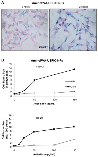
Figure 2 Time-dependent uptake of oleic acid-coated ultrasmall superparamagnetic iron oxide nanoparticles (USPIO NPs) by Caco-2 and HT-29 cells. (A) Caco-2 cells were grown to 75% confluence, then were exposed for 6 or 24 hours in complete culture medium to 20 μg/mL oleic acid-coated USPIO NPs. Then cells were stained with Prussian blue and Nuclear Red histological stains (iron blue, nucleus red, cytoplasm pink). (B) Caco-2 and HT-29 cells were grown to 90% confluence, then they were exposed for 6 hours (grey line) or 24 hours (black line) in complete culture medium to increasing concentrations of oleic acid-coated USPIO NPs and the cell-associated iron content of the cell layer was quantified using the Prussian blue reaction.
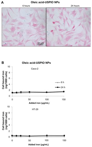
Figure 3 Cytotoxicity of aminoPVA- and oleic acid-coated USPIO NPs for colon carcinoma cells. Caco-2 cells (black line) and HT-29 cells (grey line) were exposed for 24 hours to aminoPVA-coated (A) and oleic acid-coated (B) USPIO NPs, then an MTT test was performed to ascertain cytotoxicity.
Notes: Means ± standard deviation were calculated, and statistical significance was assessed using Student’s t-test; *P < 0.05, exposed cells compared to control cells
Abbreviations: aminoPVA, polyvinylamine; USPIO NPs, ultrasmall superparamagnetic iron oxide nanoparticles.
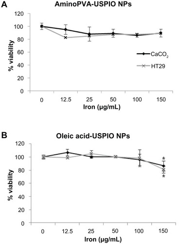
Figure 4 Cellular localization of aminoPVA-coated USPIO NPs in HT-29 cells. (A) After 6 hours of exposure, transmission electron microscopy images demonstrated that the iron oxide core of aminoPVA USPIO NPs (21 μg Fe/mL) was localized in intracellular organelles of HT-29 cells. (B) After 24 hours of exposure the iron oxide core of aminoPVA USPIO NPs (21 μg Fe/mL) was localized closer to the nucleus. (C) For the purpose of comparison, cell-associated iron was quantified by quantitative Prussian blue reaction. (D) Elemental analysis (dotted arrow) performed confirmed the presence of iron in the area defined by the black square of the transmission electron microscopy images (lower panel, right).
Abbreviations: aminoPVA, polyvinylamine; USPIO NPs, ultrasmall superparamagnetic iron oxide nanoparticles.
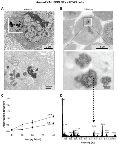
Figure 5 Polyvinylamine (aminoPVA)- and oleic acid-coated ultrasmall superparamagnetic iron oxide nanoparticles (USPIO NPs) selectively invade three-dimensional spheroids of human HT-29 and Caco-2 colon carcinoma cells. (A) Human HT-29 cells in three-dimensional spheroid cultures form well-structured spheroids after 3 days. The spheroids were exposed to aminoPVA USPIO NPs (21 μg Fe/mL) for 2 or 24 hours, recovered, fixed, then cell-associated iron was determined on histological slides by the histological Prussian blue reaction (iron, blue staining; cell cytoplasm, pink; cell nuclei, red). After 2 hours aminoPVA USPIO NPs were found in very few cells (arrow) of the first layer of the spheroids, but after 24 hours’ exposure of the spheroids they were more widely found in many cells, including in cells localized more deeply in the spheroids. (left, 100× ; right, 400× magnification). (B) Human HT-29 and Caco-2 cells in three-dimensional spheroid cultures were exposed to aminoPVA-coated USPIO NPs (21 μg iron/mL, left panels) and oleic acid-coated USPIO NPs (45 μg iron/mL, right panels) for 24 hours, recovered, fixed, then cell-associated iron (arrows) was determined on histological slides by the histological Prussian blue reaction (iron, blue staining; cell cytoplasm, pink; cell nuclei, red).
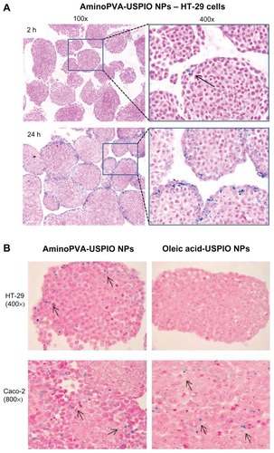
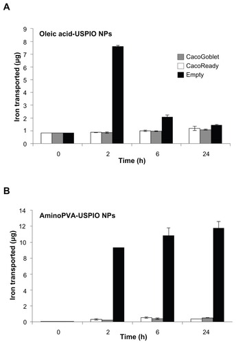
Figure 6 Transport of ultrasmall superparamagnetic iron oxide nanoparticles (USPIO NPs) by human colon cells. Transport of polyvinylamine (aminoPVA) USPIO NPs (A) and oleic acid-coated USPIO NPs (B) across CacoReady™ (white bars) and CacoGobletTM (grey bars) colon barriers and across empty membrane (black bars). The initial concentration of iron in apical chamber was 100 μg/mL (A) or 70 μg/mL (B). Iron concentration in the basal chamber was quantified with the Prussian blue reaction.
Note: The results represent noncumulated amounts of iron in the basal compartments at each time-point, without addition of USPIO NPs in the apical compartment.