Figures & data
Figure 1 (A) Western blot of preS-pulldowned proteins; (B) Western blot of SCCA1-pulldowned proteins; (C) Western blot of FTL-pulldowned proteins. For A, MBP-preS was pre-incubated with either GST-FTL or GST before mixing with amylose beads; for B, MBP-SCCA1 was pre-incubated with either GST-FTL or GST before mixing with amylose beads; for C, HA-tagged preS and SCCA1 were coexpressed with His-tagged FTL protein in HepG2 cells before immunoprecipitation by anti-His-tag antibody.
Abbreviations: MBP, maltose binding protein; GST, glutathione-S-transferase; FTL, ferritin light chain; SCCA1, squamous cell carcinoma antigen 1.

Figure 2 (A) Determination of regions on FTL for interaction with preS and SCCA1. GST-tagged FTL deletion mutant proteins, each with deletion of one of the five α-helices (a to e), were allowed to bind to MBP-preS and His-SCCA1, and absorbed with Gutathione–SepharoseTM beads. Proteins absorbed on Glutathione–Sepharose beads were subjected to Western blotting with anti-MBP antibody (detecting MBP-preS, upper), or with His-tag antisera (detecting His-SCCA1, down). (B) Verification of FTL-binding activity of N-terminal 1–11 amino acids of preS2. For the upper, pulldown assay was done by mixing GST-FTL with MBP-preS2 (lane 1) or MBP-preS2Δ (1–11) (deletion of N-terminal 1–11 amino acids of preS2, lane 2), and the proteins recovered by Glutathione–Sepharose beads were subjected to Western blot with anti-MBP antibody. For the down, pulldown assay was done by mixing GST-FTL with MBP (lane 1) or MBP-peptide (1–11) (MBP with the peptide of 11 amino acids from N-terminus of preS2, lane2), and the proteins recovered by Glutathione-Sepharose beads were subjected to Western blot with anti-MBP antibody.
Abbreviations: FTL, ferritin light chain; MBP, maltose binding protein; SCCA1, squamous cell carcinoma antigen 1; GST, glutathione-S-transferase.
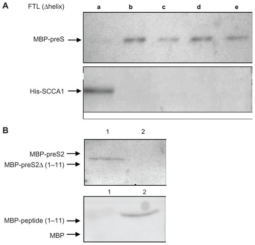
Figure 3 Effect of overexpression of SCCA1, FTL on HBV attachment in HepG2 and CHO cells. Cells were transfected with pcDNA3.1 (column 1), pcDNA3.1- SCCA1 (column 2), pcDNA3.1-FTL (column 3), or both pcDNA3.1-SCCA1 and pcDNA3.1-FTL (column 4). HBsAg dissolved in RIPA buffer was measured by ELISA assay to quantify the HBV attachment experiment.
Abbreviations: CHO, Chinese hamster ovary; SCCA1, squamous cell carcinoma antigen 1; FTL, ferritin light chain; HBV, hepatitis B virus; HBsAg, hepatitis B virus surface protein antigens; RIPA, radio immunoprecipitation assay; ELISA, enzymelinked immunosorbent assay.
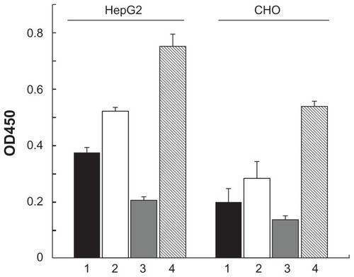
Figure 4 Coexpression of human SCCA1 and FTL in mouse liver enhances HBV replication. Plasmids pcDNA3.1 (column 1 in A; a in B), pcDNA3.1-SCCA1 (column 2 in A; b in B), pcDNA3.1-FTL (column 3 in A; c in B), or both of pcDNA3.1-SCCA1 and pcDNA3.1-FTL (column 4 in A; d in B) were introduced into mouse liver via hydrodynamic injection through tail vein. (A) Serum level of HBsAg determined 24 hours post virus challenge. (B) Immunohistochemical staining of HBcAg 48 hours post virus challenge.
Abbreviations: SCCA1, squamous cell carcinoma antigen 1; FTL, ferritin light chain; HBV, hepatitis B virus; HBsAg, hepatitis B virus surface protein antigens; HBcAg, hepatitis B virus core protein antigens.
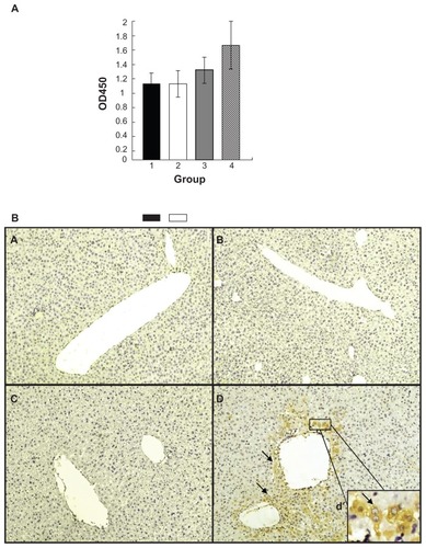
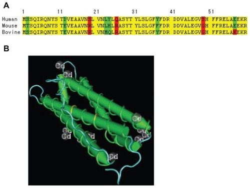
Figure 5 (A) Comparison of the FTL N-terminal amino acids sequence of human, mouse and bovine. (B) Illustration of the position of S19R and Q26R on 3D structure.
Abbreviation: FTL, ferritin light chain.