Figures & data
Table 1 Physicochemical characteristics of nanoformulations of antiretroviral drugs
Figure 1 Scanning electron microscopy (SEM) images of nanoART morphology.
Notes: SEM analysis (magnification, 15,000×) of nanoformulated IDV, RTV, ATV, and EFV produced by homogenization, wet-milling, and sonication on top of a 0.2 μm polycarbonate filtration membrane. Scale bar equals 10 μm for IDV and 3 μm for RTV, ATV and EFV. Wet milling and homogenization show crystalline nanoparticles while sonicated formulations are seen as spherical nanoparticles.
Abbreviations: IDV, indinavir; RTV, ritonavir; ATV, atazanavir; EFV, efavirenz.
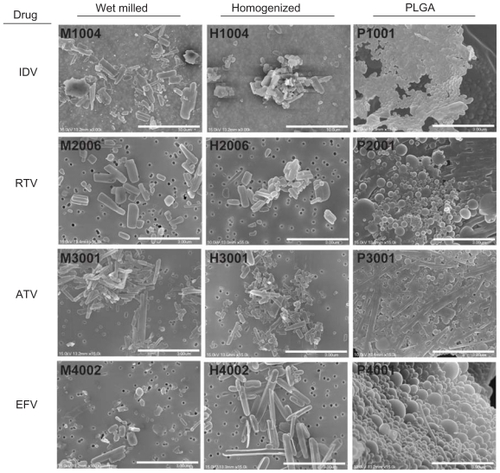
Figure 2 Macrophage uptake, retention and release of nanoART.
Notes: Time courses of monocyte-derived macrophage (MDM) uptake of IDV, RTV, ATV, and EFV nanoparticles manufactured by wet-milling, homogenization or sonication over a period of 8 hours and cell retention and release over a period of 15 days after treatment. Data represent the mean ± SEM for n = 3 determinations per time.
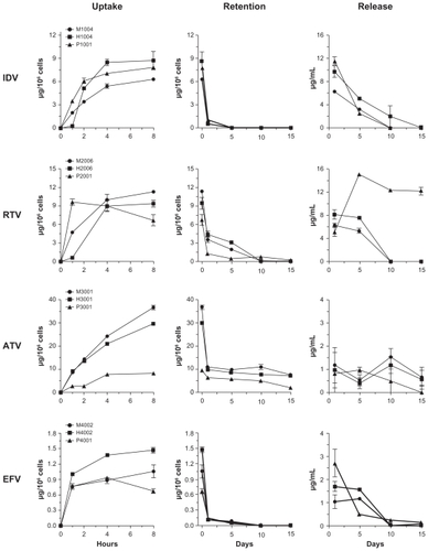
Figure 3 Comparison of antiretroviral effects of various formulations of IDV, RTV, ATV, and EFV as measured by reverse transcriptase (RT) activity.
Notes: Monocyte-derived macrophages (MDM) were treated with 100 μM of nanoparticles for 8 hours. MDM were infected with HIV-1ADA on days 1, 5, 10, and 15 post-loading and cultured for 10 days after infection. Medium was removed and RT activity was measured by 3H-TTP incorporation. Data are normalized to activity in HIV-1 infected untreated cells cultured in parallel. Data represent mean ± SEM for n = 4 determinations per treatment.

Figure 4 Antiretroviral efficacy of nanoART as determined by HIV-1p24 antigen staining.
Notes: Monocyte-derived macrophages (MDM) were loaded for 8 hours with IDV, RTV, ATV and EFV nanoparticles manufactured by (A) wet milling and homogenization or (B) sonication. MDM were infected with HIV-1ADA on days 1, 5, 10, and 15 post-loading and cultured for an additional 10 days. MDM were fixed and immunostained for expression of p24. Untreated, uninfected MDM served as negative controls (control), while MDM exposed to HIV-1ADA but not treated with nanoART, served as positive controls (+HIV). Expression of viral p24 antigen was visualized by DAB chromogen (brown). Images are representative of n = 4 determinations per treatment. (Magnification, 200×).
Abbreviations: IDV, indinavir; RTV, ritonavir; ATV, atazanavir; EFV, efavirenz.
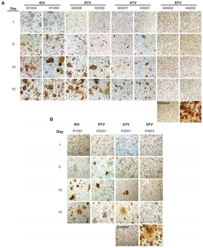
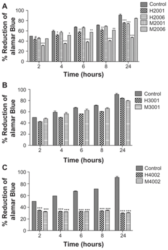
Figure 5 Effect of nanoART on macrophage viability.
Notes: Monocyte-derived macrophages (MDM) were loaded for 12 hours with (A) four nanoformulations of RTV (H2001, H2006, M2001, M2006), (B) two nanoformulations of ATV (H3001 and M3001), or (C) two nanoformulations of EFV (H4002 and M4002) at 0.1 mM. Controls consisted of untreated cells. Following drug loading, toxicity was assessed over 24 hours by alamarBlueTM assay. For each experimental condition, n = 3. Figure shown is representative of two independent experiments. Significant differences are indicated: #(P < 0.05), *(P < 0.01), ** (P < 0.001), ***(P < 0.0001).