Figures & data
Figure 1 Scanning electron microscopy/energy dispersive X-ray spectroscopy (EDS) mapping of various porous gelatin scaffolds modified with chondroitin-4-sulfate (GCS000, GCS005, GCS025, and GCS125): (A) scanning electron microscopy images, (B) EDS mapping of sulfur, and (C) EDS spectrogram.
Notes: Scale bars are 50 μm; scaffold groups labeled according to chondroitin-4-sulfate concentration used (0%, 0.05%, 0.25%, or 1.25% (w/v)): GCS000, GCS005, GCS025, and GCS125.
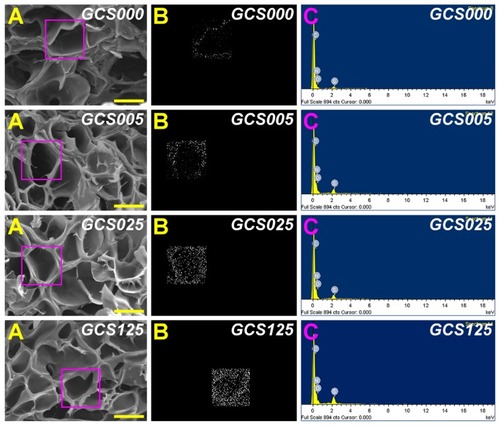
Figure 2 (A) Fourier transform infrared spectroscopy spectra of chondroitin-4-sulfate (CS) and the gelatin samples modified with varying concentrations of CS (0%–1.25% (w/v)); (B) the absorption peak ratios of the S=O stretching to N–H stretching bands (A1228/A3294) and C–O stretching to N–H stretching bands (A1403/A3294) for the gelatin samples modified with varying concentrations of CS (GCS000, GCS005, GCS025, and GCS125).
Notes: Values are mean plus or minus standard deviation (n = 3); *P < 0.05 versus all groups (compared only within A1228/A3294 or A1403/A3294 groups); scaffold groups labeled according to CS concentration used (0%, 0.05%, 0.25%, or 1.25% (w/v)): GCS000, GCS005, GCS025, and GCS125.
Abbreviation: A/A, absorbance/absorbance.
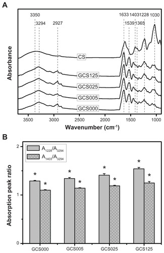
Figure 3 The chondroitin-4-sulfate (CS) content (determined by dimethylmethylene blue assay) of the gelatin scaffolds modified with varying concentrations of CS (0%–1.25% (w/v)).
Notes: Values are mean plus or minus standard deviation (n = 4); *P < 0.05 versus all groups.
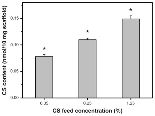
Figure 4 Equilibrium water content of various porous gelatin scaffolds modified with concentrations of chondroitin-4-sulfate (GCS000, GCS005, GCS025, and GCS125).
Notes: Values are mean plus or minus standard deviation (n = 6); *P < 0.05 versus all groups; scaffold groups labeled according to chondroitin-4-sulfate concentration used (0%, 0.05%, 0.25%, or 1.25% (w/v)): GCS000, GCS005, GCS025, and GCS125.
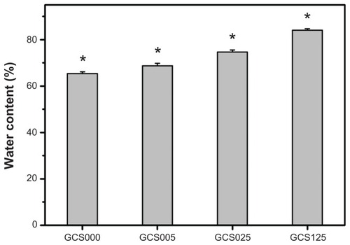
Figure 5 Optical transmittance of various porous gelatin scaffolds (GCS000, GCS005, GCS025, and GCS125) modified with concentrations of chondroitin-4-sulfate.
Notes: Scaffold groups labeled according to chondroitin-4-sulfate concentration used (0%, 0.05%, 0.25%, or 1.25% (w/v)): GCS000, GCS005, GCS025, and GCS125.
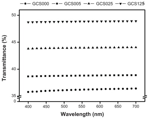
Figure 6 Young’s modulus of various porous gelatin scaffolds (GCS000, GCS005, GCS025, and GCS125) modified with concentrations of chondroitin-4-sulfate.
Notes: Values are mean plus or minus standard deviation (n = 8); *P < 0.05 versus all groups; #P < 0.05 versus GCS025 and GCS125 groups; scaffold groups labeled according to chondroitin-4-sulfate concentration used (0%, 0.05%, 0.25%, or 1.25% (w/v)): GCS000, GCS005, GCS025, and GCS125.
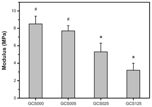
Figure 7 Time course of the percentage of weight remaining for various chondroitin-4-sulfate-modified porous gelatin scaffolds (GCS000, GCS005, GCS025, and GCS125) after incubation at 34°C in a balanced salt solution containing collagenase.
Notes: An asterisk indicates statistically significant differences (*P < 0.05; n = 5) for the mean value of weight remaining compared with value at previous time point; #P < 0.05 versus all groups; +P < 0.05 versus GCS025 and GCS125 groups; ^P < 0.05 versus GCS000 and GCS005 groups; –P < 0.05 versus GCS000 and GCS125 groups (compared only within each time point group); scaffold groups labeled according to chondroitin-4-sulfate concentration used (0%, 0.05%, 0.25%, or 1.25% (w/v)): GCS000, GCS005, GCS025, and GCS125.
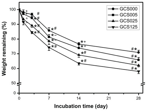
Figure 8 Concentration of glucose permeated through various chondroitin-4-sulfate-modified porous gelatin scaffolds (GCS000, GCS005, GCS025, and GCS125) at 34°C.
Notes: Values are mean plus or minus standard deviation (n = 5); *P < 0.05 versus all groups; scaffold groups labeled according to chondroitin-4-sulfate concentration used (0%, 0.05%, 0.25%, or 1.25% (w/v)): GCS000, GCS005, GCS025, and GCS125.
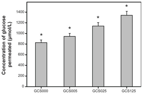
Figure 9 (A) Phase contrast micrographs of rabbit corneal keratocyte cultures incubated for 2 days at 37°C with extract medium conditioned with various chondroitin-4-sulfate-modified porous gelatin scaffolds (control, GCS000, GCS005, GCS025, and GCS125); (B) gene expression of interleukin-6 (IL-6) in rabbit corneal keratocytes incubated with extract medium conditioned with various chondroitin-4-sulfate-modified porous gelatin scaffolds for 2 days, measured by real-time reverse transcription polymerase chain reaction.
Notes: Scale bars, 100 μm; glyceraldehyde-3-phosphate dehydrogenase was used for normalization; data in the experimental groups are percentages relative to that of control groups (without materials); values are mean plus or minus standard deviation (n = 3); scaffold groups labeled according to chondroitin-4-sulfate concentration used (0%, 0.05%, 0.25%, or 1.25% (w/v)): GCS000, GCS005, GCS025, and GCS125.
Abbreviation: mRNA, messenger RNA.
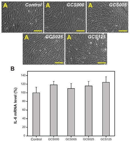
Figure 10 Total cell number on various chondroitin-4-sulfate-modified porous gelatin scaffolds (GCS000, GCS005, GCS025, and GCS125). after rabbit corneal keratocyte seeding for 1, 3, and 5 days (d1, d3, and d5, respectively).
Notes: An asterisk indicates statistically significant differences (*P < 0.05; n = 4) for the mean value of total cell number compared with value at previous time point; #P < 0.05 versus all groups; +P < 0.05 versus GCS025 and GCS125 groups (compared only within each time point group); scaffold groups labeled according to chondroitin-4-sulfate concentration used (0%, 0.05%, 0.25%, or 1.25% (w/v)): GCS000, GCS005, GCS025, and GCS125.
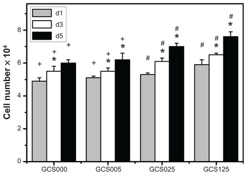
Figure 11 Extracellular matrix production capacity of rabbit corneal keratocytes at days 1, 3, and 5 (d1, d3, and d5, respectively) after plating on various chondroitin-4-sulfate-modified porous gelatin scaffolds (GCS000, GCS005, GCS025, and GCS125): (A) collagen content and (B) glycosaminoglycan (GAG) content.
Notes: An asterisk indicates statistically significant differences (*P < 0.05; n = 3) for the mean value of extracellular matrix content compared with value at previous time point; #P < 0.05 versus all groups (compared only within each time point group); scaffold groups labeled according to chondroitin-4-sulfate concentration used (0%, 0.05%, 0.25%, or 1.25% (w/v)): GCS000, GCS005, GCS025, and GCS125.
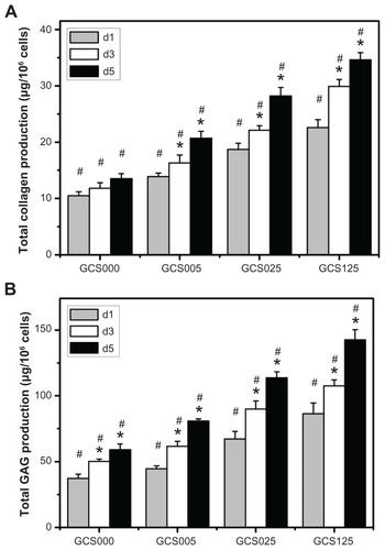
Figure 12 Scanning electron microscopy images of (A and B) the constructs prepared from 5 days’ cultivation of the rabbit corneal keratocytes on scaffold samples and (C and D) the cell-free scaffolds (large arrow indicates cell, fine arrow indicates extracellular matrix). Groups are labeled according to chondroitin-4-sulfate concentration used: (A and C) GCS000 (0% (w/v)) and (B and D) GCS025 (0.25% (w/v)). (E) Gene expression level of keratocan and biglycan in rabbit corneal keratocytes grown on tissue culture polystyrene and scaffold samples GCS000 and GCS025 for 5 days by real-time reverse transcription polymerase chain reaction.
Notes: Scale bars, 20 μm; glyceraldehyde-3-phosphate dehydrogenase was used for normalization; data in the experimental groups are percentages relative to that of tissue culture polystyrene groups; an asterisk indicates statistically significant differences (*P < 0.05; n = 3) as compared with the GCS000 groups.
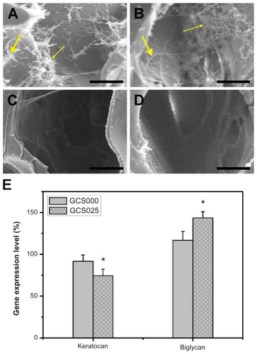
Figure S1 Cross-linking index of porous gelatin scaffolds treated with 1-ethyl-3-(3-dimethyl aminopropyl) carbodiimide hydrochloride/N-hydroxysuccinimide as a function of cross-linking time.
Notes: An asterisk indicates statistically significant differences (*P < 0.05; n = 5) for the mean value of cross-linking index compared with the value at the previous time point.
Abbreviations: EDC, 1-ethyl-3-(3-dimethyl aminopropyl) carbodiimide hydrochloride; NHS, N-hydroxysuccinimide.
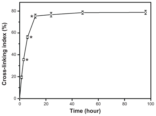
Figure S2 Pore size of various porous gelatin scaffolds modified with chondroitin-4-sulfate.
Notes: Values are mean plus or minus standard deviation (n = 3); scaffold groups labeled according to chondroitin-4-sulfate concentration used (0%, 0.05%, 0.25%, or 1.25% (w/v)): GCS000, GCS005, GCS025, and GCS125.
Abbreviation: CS, chondroitin-4-sulfate.
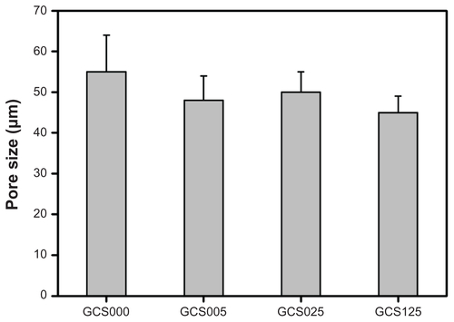
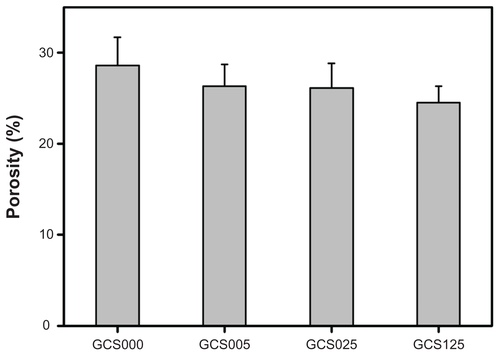
Figure S3 Porosity of various porous gelatin scaffolds modified with chondroitin-4-sulfate.
Notes: Values are mean plus or minus standard deviation (n = 5); scaffold groups labeled according to chondroitin-4-sulfate concentration used (0%, 0.05%, 0.25%, or 1.25% (w/v)): GCS000, GCS005, GCS025, and GCS125.
Abbreviation: CS, chondroitin-4-sulfate.