Figures & data
Figure 1 Characterization of silver nanoparticles. (A) Silver nanoparticles showing spherical, monodispersed particles (scale bar, 50 nm). The inset shows one single particle of silver (scale bar, 5 nm). (B) Particle size distribution showing preponderance of particles in the size range of 10–15 nm. (C) Electron diffraction pattern of nanoparticles showing various crystallographic planes. (D) Optical spectra of silver before (1) and after reduction (2). The inset shows the corresponding change in color.
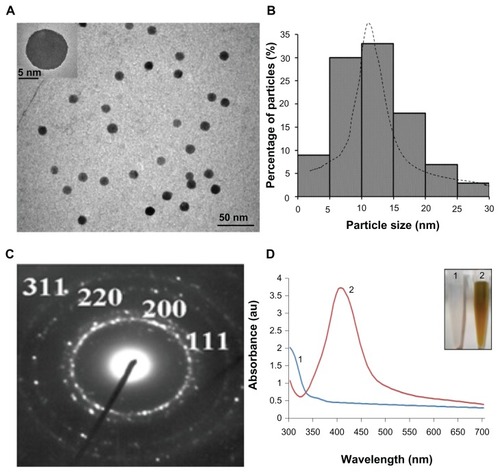
Table 1 Brugia malayi microfilariae were incubated with silver nanoparticles at varying concentrations
Table 2 Percentage inhibition in poly(adenosine diphosphateribose) polymerase activity in the microfilariae treated with different reagents as indicated compared with control (Roswell Park Memorial Institute medium only)
Figure 2 Ethidium bromide/acridine orange differential staining of microfilarial forms for the detection of apoptosis. Untreated (A) and gold nanoparticles preincubated (D) nuclei showed green staining due to acridine orange permeation, while organisms treated with silver nanoparticles (B) and staurosporine (C) appeared orange-yellow due to ethidium bromide, suggesting loss of integrity of surface membrane of the parasite shown.
Note: Data are representative of three different experiments.
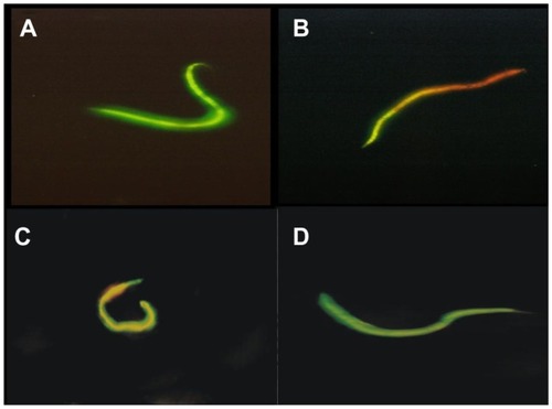
Figure 3 Scanning electron micrographs of (A) untreated control parasite in Roswell Park Memorial Institute medium; (B) microfilariae treated with staurosporine (0.5 μM); (C) microfilariae treated with silver nanoparticles (50 μM), and (D) microfilariae treated with gold nanoparticles (50 μM).
Note: Data are representative of three different experiments.
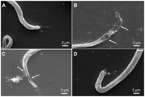
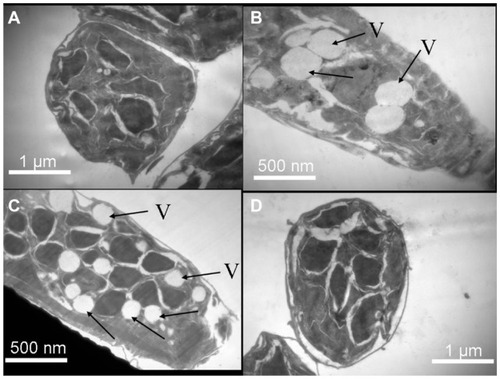
Figure 4 Transmission electron micrographs through sections of (A) untreated control parasite in Roswell Park Memorial Institute medium; (B) microfilariae treated with staurosporine (0.5 μM); (C) microfilariae treated with silver nanoparticles (50 μM); and (D) microfilariae treated with gold nanoparticles (50 μM).
Notes: Arrows point to the vacuoles; data are representative of three different experiments.
Abbreviation: V, vacuoles.