Figures & data
Table 1 Biophysical characterization of lipophilic carrier formulations and free GP. The results are presented as the mean ± standard deviation
Figure 1 Tissue residence of free GP, GPNP, and GPNP-PEG at 6 and 24 hours.
Notes: Data are presented as the ppb Au/g tissue ± standard deviation (*P < 0.05, n = 3).
Abbreviations: GP, gold porphyrin; GPNP, gold porphyrin nanoparticles; GPNP-PEG, gold porphyrin nanoparticles surface-coated with PEG.
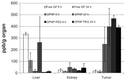
Figure 2 (A) Dose-dependent antitumor activity against N2 A for GP and camptothecin with or without the lipophilic carrier formulation (n = 4, *P < 0.05 when comparing nanoparticle formulation with free GP or camptothecin). (B) Comparison of surface-coated lipophilic nanoparticle carrier with different molecular weights (750 Da and 2000 Da) of PEG.
Notes: Results are presented as the mean ± standard deviation (n = 4, *P < 0.05 for C-PEG 750 or C-PEG 2000 in comparison with C).
Abbreviations: C, camptothecin; GP, gold porphyrin; GPNP, gold porphyrin nanoparticles; GPNP-PEG, gold porphyrin nanoparticles surface-coated with PEG.

Figure 3 Tumor volume measurements after N2A-bearing A/J mice had received intraperitoneal injections of GPNP-PEG 3, 4, or 5 mg/kg.
Notes: n = 5–6, *P < 0.05.
Abbreviation: GPNP-PEG, gold porphyrin nanoparticles surface-coated with PEG.
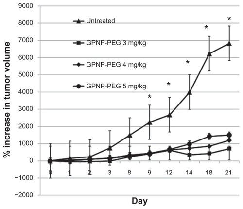
Figure 4 Apoptotic activity of (A) GP formulations and (B) camptothecin formulations using Tunel assay in neuroblastoma tissue harvested at 21 days after treatment.
Notes: Green represents positive apoptotic cells and blue represents the DAPI nuclei stain. Magnification 100× (left column) or 200× (right column); n = 5, *P < 0.05 for 4 mg/kg or 5 mg/kg of GPNP-PEG in comparison with untreated controls in Figure 4A.
Abbreviations: GP, gold porphyrin; GPNP-PEG, gold porphyrin nanoparticles surface-coated with PEG.
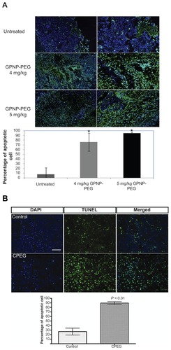
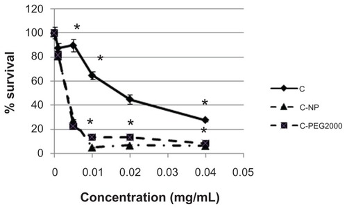
Figure 5 Dose-dependent antitumor activity against D54.
Notes: Results are presented as the mean ± standard deviation (n = 4, *P < 0.05 for C-NP or C-PEG 2000 compared with C).