Figures & data
Figure 1 General reaction scheme for synthesis of the mannosylated polyethylene glycol-phosphatidylethanolamine ligand.
Abbreviations: Boc, butyl carbonyl; PE, phosphatidylethanolamine; PEG, polyethylene glycol; DMSO, dimethyl sulfoxide; TEA, triethylamine.
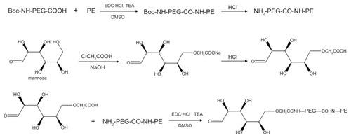
Figure 2 Preparation and modification of liposome-pEGFP complexes.
Abbreviations: M-PEG-PE, mannosylated polyethylene glycol-phosphatidylethanolamine; Lipo, Lipofectamine 2000; pEGFP, green fluorescence protein plasmid.
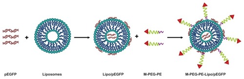
Figure 3 Transmission electron microscopic images of Lipofectamine-green fluorescence protein plasmid (A), mannosylated polyethylene glycol-phosphatidylethanolamine- Lipofectamine-green fluorescence protein plasmid (B and C).
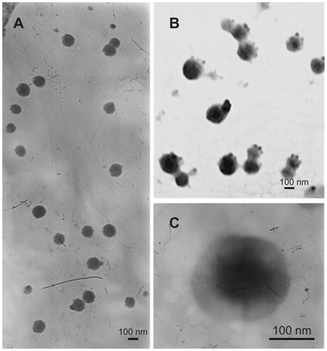
Table 1 Particle size and zeta potential of liposomes (mean ± standard deviation, n = 3)
Figure 4 Optimization of the M-PEG-PE modification rate.
Abbreviations: M-PEG-PE, mannosylated polyethylene glycol-phosphatidylethanolamine; Lipo, Lipofectamine™; pEGFP, green fluorescence protein plasmid.

Figure 5 Turbidity of Lipofectamine™ 2000, Lipo-pEGFP, and M-PEG-PE-Lipo-pEGFP at different time intervals.
Abbreviations: M-PEG-PE, mannosylated polyethylene glycol-phosphatidylethanolamine; Lipo, Lipofectamine™; pEGFP, green fluorescence protein plasmid.
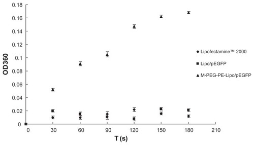
Figure 6 Cell viability of M-PEG-PE-Lipo/pEGFP and Lipo/pEGFP.
Abbreviations: M-PEG-PE, mannosylated polyethylene glycol-phosphatidylethanolamine; Lipo, Lipofectamine™; pEGFP, green fluorescence protein plasmid.
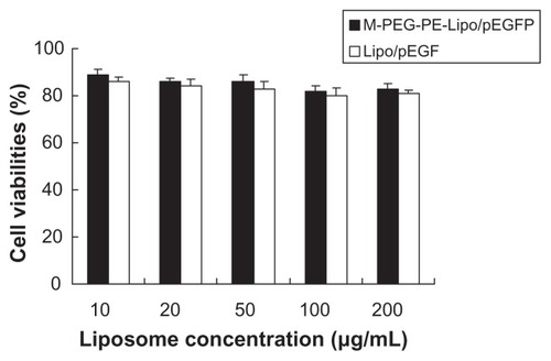
Figure 7 Fluorescent images and flow cytometry analysis of Kupffer cells transfected with naked pEGFP; (A), Lipo/pEGFP (B), and M-PEG-PE-Lipo/pEGFP (C) at 24, 48, and 72 hours following transfection, respectively.
Abbreviations: M-PEG-PE, mannosylated polyethylene glycol-phosphatidylethanolamine; Lipo, Lipofectamine™; pEGFP, green fluorescence protein plasmid.
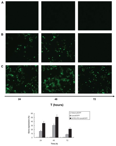
Figure 8 Flow cytometry analysis of Kupffer cells transfected by naked pEGFP (A), Lipo-pEGFP (B), and M-PEG-PE-Lipo-pEGFP (C) in vivo. Gene expression was examined 48 hours following injection.
Abbreviations: M-PEG-PE, mannosylated polyethylene glycol-phosphatidylethanolamine; Lipo, Lipofectamine™; pEGFP, green fluorescence protein plasmid.
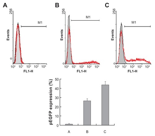
The data in the quadrantal rule diagram for the in vivo study are as follows
Data on dose optimization are as follows (gene expression was analyzed at 48 hours after intravenous injection)