Figures & data
Table 1 Composition and properties of prepared liposomal doxorubicin and AP-1 liposomal doxorubicin
Figure 1 (A) Schematic representation of the AP-1-conjugated liposome. Liposomes were prepared containing maleimide functional polyethylene glycol chains. Maleimide was used to attach the AP-1 peptide through the thiol group on a cysteine. (B) Flow cytometric detection of the cell-surface interleukin-4 receptor on cloned human GBM8401-luc cells. (C) Linearity of measured bioluminescence versus GBM8401-luc cell number. GBM8401-luc cells were plated into 96-well dishes in triplicate in various numbers, and were then imaged by the in vivo imaging system. A strong correlation (R2 = 0.996) was observed between luciferase activity and cell numbers. (D) A luciferase image of the plated GBM8401-luc cells.
Abbreviations: IL-4R, interleukin-4 receptor; Lipo-Dox, liposomal doxorubicin.
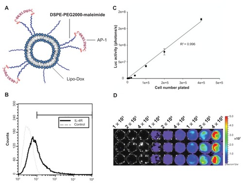
Figure 2 Influence of the concentration of liposomal doxorubicin on tumor progression and bodyweight. (A) Control mice are indicated by ●. Tumor-bearing mice were treated on day 5 with different doses of liposomal doxorubicin (▾, 5 mg/kg; ○, 10 mg/kg). Each point consisted of three mice. *Significant difference in control mice over the three days following cell numbers on day 5 after implantation. There was a significant difference between the tumors treated with liposomal doxorubicin at doses of 5 mg/kg or 10 mg/kg and control tumors on day 12 after implantation. (B) Body weight change (relative to day 1) of tumor-bearing mice treated with different doses of liposomal doxorubicin.
Notes: *P < 0.05; #P < 0.05; n = 3.
Abbreviation: Lipo-Dox, liposomal doxorubicin.
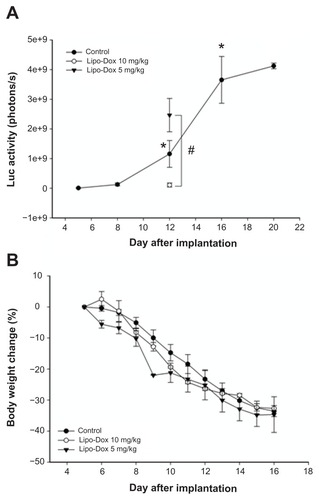
Figure 3 (A) Concentration of doxorubicin delivered to the brain as a function of acoustic power. Compared with the nonsonicated normal brain, there was a significant difference for sonicated brains at 2.86 W or 4.29 W. **,#/##Significant difference compared with the nonsonicated brain and the brain sonicated at 1.43 W, respectively. The concentrations of interleukin-4 receptor protein and vascular endothelial growth factor protein were evaluated in the brain tumor and the neighboring brain, respectively. There was no significant difference in protein expression of the interleukin-4 receptor (B) or vascular endothelial growth factor (C) after pulsed HIFU exposure.
Notes: #P < 0.05; **P < 0.01; ##P < 0.01; n = 3).
Abbreviations: Lipo-Dox, liposomal doxorubicin; HIFU, high-intensity focused ultrasound; VEGF, vascular endothelial growth factor.
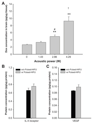
Figure 4 (A) Measurements of untargeted liposomal doxorubicin and AP-1-conjugated liposomal doxorubicin in the contralateral normal brain and the brain tumor without and with repeated sonication. Compared with the contralateral normal brain, there was a significant difference between the control tumors and the repeatedly sonicated tumors with the two drugs. The concentrations of doxorubicin in the brain tumors after repeated sonication were significantly higher than in brain tumors without sonication. *,***,###Significant difference compared with contralateral normal brain and the nonsonicated brain tumor, respectively. (B) Derived tumor-to-contralateral brain ratios without and with repeated sonication after drug administration. The values of the ratios are significantly elevated after application of repeated sonication.
Notes: *P < 0.05; ***P < 0.001; ###P < 0.001; n = 3.
Abbreviations: AP-1, atherosclerotic plaque-specific peptide-1; Lipo-Dox, liposomal doxorubicin; HIFU, high-intensity focused ultrasound.
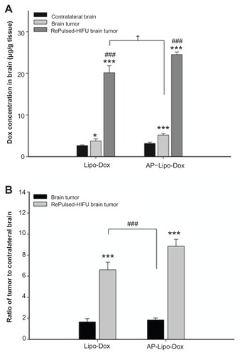
Figure 5 Longitudinal bioluminescence imaging of the brain tumors was monitored from 5 to 16 days after implantation. Firefly luciferase-labeled GBM8401 cells had been implanted into the left forebrain of NOD-scid mice which received no treatment (control), post-treatment with repeated pulsed HIFU after liposomal doxorubicin on day 5, or post-treatment with repeated pulsed HIFU after AP-1 liposomal doxorubicin injection on day 5.
Abbreviations: AP-1, atherosclerotic plaque-specific peptide-1; Lipo-Dox, liposomal doxorubicin; HIFU, high-intensity focused ultrasound.
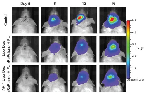
Figure 6 (A) Analysis of increases in tumor size (relative to day 5) is based on data obtained from bioluminescence images in . Compared with the control tumor mice, * and # denote a significant difference for untargeted liposomal doxorubicin with repeated sonication and AP-1 liposomal doxorubicin with repeated sonication, respectively. (B) Bodyweight change (relative to day 5) of tumor-bearing mice treated by untargeted liposomal doxorubicin with repeated sonication and AP-1 liposomal doxorubicin with repeated sonication.
Notes: *P < 0.05; #P < 0.05.
Abbreviations: AP-1, atherosclerotic plaque-specific peptide-1; Lipo-Dox, liposomal doxorubicin; HIFU, high-intensity focused ultrasound.
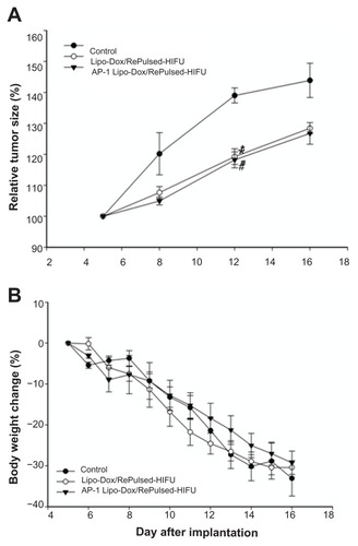
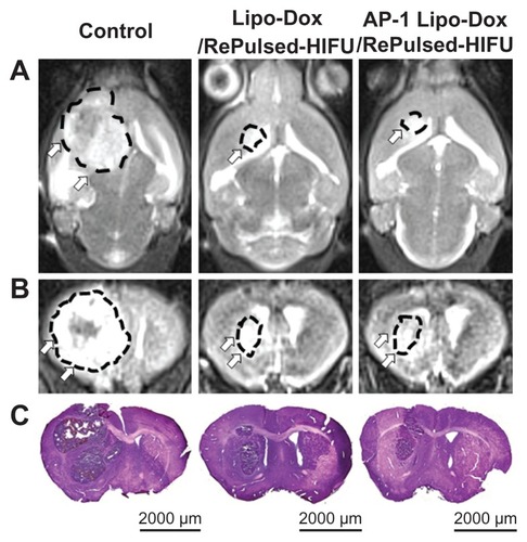
Figure 7 Twelve days after tumor implantation, three mice each from the control group, the untargeted liposomal doxorubicin with repeated sonication group, and the AP-1 liposomal doxorubicin with repeated sonication group were imaged by T2-weighted magnetic resonance imaging (A, coronal view; B, axial view), and then sacrificed for hematoxylin and eosin (C) histological examination (scale bar, 2000 μm).
Abbreviations: AP-1, atherosclerotic plaque-specific peptide-1; Lipo-Dox, liposomal doxorubicin; HIFU, high-intensity focused ultrasound.