Figures & data
Figure 1 Illustration of D-USPIO and DM-USPIO.
Note: DM-USPIO was prepared in-house by adding alkali-treated mannan to D-USPIO.
Abbreviations: D-USPIO, dextran-coated ultrasmall superparamagnetic iron oxide; DM-USPIO, mannan–dextran-coated ultrasmall superparamagnetic iron oxide.
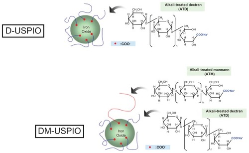
Figure 2 In vivo MRA of the thoracic aorta of WHHL rabbits, (A) before, and (B) after the administration of D-USPIO, and (C) before, and (D) after the administration of DM-USPIO.
Notes: Irregularities, seen as spotty signal voids in the aortic wall, are present on the images obtained after administration of D- or DM-USPIO. The graph compares pre- and post-administration SNR measurements in the ROI in the aortic wall of WHHL rabbits subjected to in vivo MRI (n = 6), after injection with D- or DM-USPIO (n = 3, respectively). The SNR values were significantly lower on all post-contrast images. In rabbits injected with DM-USPIO, the difference between the pre- and post-administration SNR values was significantly greater than in those treated with D-USPIO *P < 0.05.
Abbreviations: D-USPIO, dextran-coated ultrasmall superparamagnetic iron oxide; DM-USPIO, mannan–dextran-coated ultrasmall superparamagnetic iron oxide; MRA, magnetic resonance angiography; SNR, signal-to-noise ratio; ROI, region of interest; MRI, magnetic resonance imaging; WHHL, Watanabe heritable hyperlipidemic.
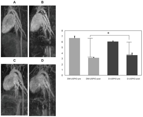
Figure 3 Graph showing the SNR of the aortic wall specimens on in vitro MRI scans.
Notes: Aortic specimens from WHHL rabbits (A) Controls; (B) dose 1, (C) dose 2, (D) dose 3 of D-USPIO; (E) dose 1, (F) dose 2, (G) dose 3 of DM-USPIO (dose 1: n = 1; dose 2: n = 1; dose 3: n = 3). Compared to the controls, the signal from the aortic wall decreased in rabbits injected with D- or DM-USPIO, in a dose-dependent manner. At dose 3, the SNR tended to be lower (*P < 0.1) in rabbits treated with DM-USPIO. The SNR value obtained from three ROIs on three different aortic images was significantly lower (**P < 0.05) in the rabbit treated with dose 2 DM-USPIO than in the rabbit treated with an equivalent dose of D-USPIO. There was no significant difference at dose 1 (P > 0.1).
Abbreviations: D-USPIO, dextran-coated ultrasmall superparamagnetic iron oxide; DM-USPIO, mannan–dextran-coated ultrasmall superparamagnetic iron oxide; SNR, signal-to-noise ratio; ROI, region of interest.
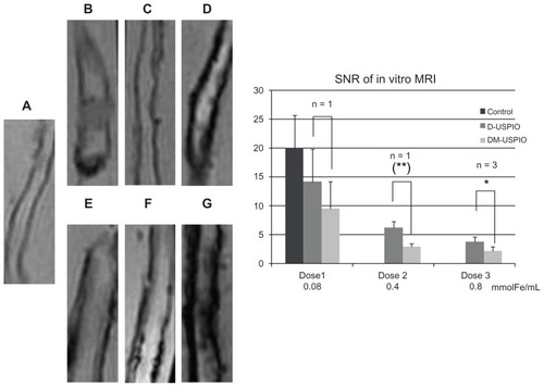
Figure 4 Prussian blue staining of histopathological sections.
Notes: (A) dose 1, (B) dose 2, (C) dose 3 of D-USPIO; (D) dose 1, (E) dose 2, (F) dose 3 of DM-USPIO. There was a marked iron uptake in the arterial walls of all rabbits injected with either D- or DM-USPIO. The Prussian blue-stained area increased in both the D- and DM-USPIO group in a dose-dependent manner.
Abbreviations: D-USPIO, dextran-coated ultrasmall superparamagnetic iron oxide; DM-USPIO, mannan–dextran-coated ultrasmall superparamagnetic iron oxide.
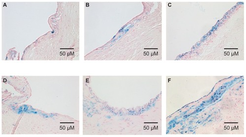
Figure 5 (A) Prussian blue and (B) immunohistochemical (RAM11) staining of histopathological sections from WHHL rabbits injected with DM-USPIO at dose 3 (0.8 mmol Fe/Kg).
Note: The localization of iron and macrophages was correlated.
Abbreviations: DM-USPIO, mannan–dextran-coated ultrasmall superparamagnetic iron oxide; WHHL, Watanabe heritable hyperlipidemic.
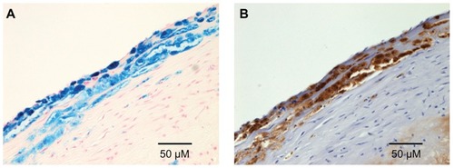
Figure 6 Iron-positive areas in the aortic wall of rabbits injected with D- or DM-USPIO.
Notes: At dose 3 (0.8 mmol Fe/Kg), the iron-positive areas were significantly larger in rabbits treated with DM- than in those treated with D-USPIO (*P < 0.05). The areas were significantly larger in different sections of the aorta from the rabbit injected with dose 1 or dose 2 DM- than in that treated with an equivalent dose of D-USPIO (*P < 0.05).
Abbreviations: D-USPIO, dextran-coated ultrasmall superparamagnetic iron oxide; DM-USPIO, mannan–dextran-coated ultrasmall superparamagnetic iron oxide.
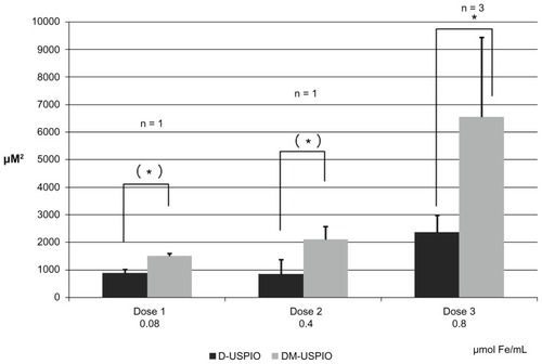
Figure 7 Iron content per unit weight of aortic specimen from rabbits injected with D- or DM-USPIO at dose 3 (0.8 mmol Fe/Kg).
Note: There was no significant difference between the two groups.
Abbreviations: D-USPIO, dextran-coated ultrasmall superparamagnetic iron oxide; DM-USPIO, mannan–dextran-coated ultrasmall superparamagnetic iron oxide.
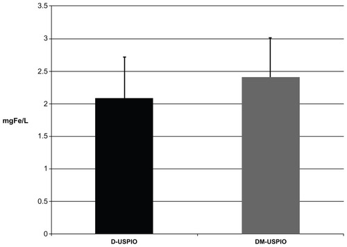
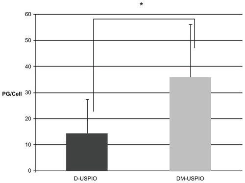
Figure 8 Iron uptake in cultured J774.1 macrophage cells, measured by atomic absorption photometry.
Note: The amount of intracellular iron was significantly higher in cells treated with DM-USPIO than with D-USPIO (*P < 0.05).
Abbreviations: D-USPIO, dextran-coated ultrasmall superparamagnetic iron oxide; DM-USPIO, mannan–dextran-coated ultrasmall superparamagnetic iron oxide.