Figures & data
Figure 1 Experimental arrangement. The acoustic beam from the ultrasound transducer to sonicate mouse ear tumors was guided by a cone filled with degassed water. A 3-mm-thick ultrasound transmission gel was mantled over the ear skin and the acoustic beam was targeted on the tumor. The ultrasound beam was circularly scanned during sonication.

Figure 2 Effects of ultrasound (US) sonication with microbubbles (MBs) on tumor growth for early stage tumors treated with 10 mg/kg of anticancer nanodrug. The arrows indicate the schedule for treatments. The figure shows the tumor growth responses for an initial size of treated tumors of about 15 mm3 with different conditions: control, PLD alone, PLD + MBs + US, US alone, and MBs + US.
Notes: *P < 0.05; **P < 0.01 (Mann–Whitney U test); for each group, mean ± standard deviation.
Abbreviations: US, ultrasound; MBs, microbubbles; PLD, pegylated liposomal doxorubicin.
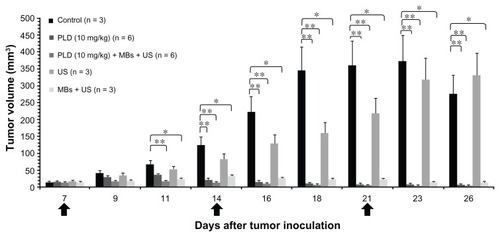
Figure 3 Effects of focused US sonication with MBs on tumor growth for medium-stage tumors treated with 6 mg/kg. (A) Tumor growth responses for an initial size of treated tumors of about 50 mm3 with different conditions: control, PLD alone, PLD + MBs + US, US alone, and MBs + US. The arrows indicate the schedule for treatments. (B) Photomicrographs of mouse-ear tumors (control, PLD alone, and PLD + MBs + US) on days 10, 16, and 26 after tumor inoculation (magnification: 10×). (C) Blood perfusion measurements of tumors before treatment on days 10 and 17, for the control, PLD, and PLD + MBs + US groups.
Notes: Mean ± SD; *P < 0.05, **P < 0.01 (Mann–Whitney U test).
Abbreviations: US, ultrasound; MBs, microbubbles; PLD, pegylated liposomal doxorubicin.
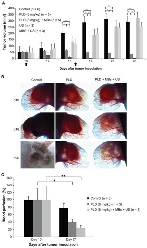
Figure 4 H&E staining of tissue sections for the ear tumors after a sequence of treatments with MBs + US, PLD alone, and PLD + MBs + US.
Notes: Magnifications 20×, 100×, 200×, and 400× (yellow square).
Abbreviations: H&E, hematoxylin and eosin; MBs, microbubbles; US, ultrasound; PLD, pegylated liposomal doxorubicin.
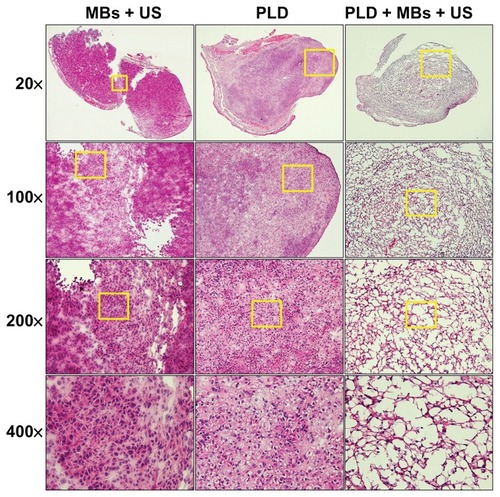
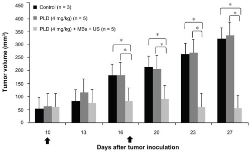
Figure 5 Tumor growth response of medium-staged tumors treated with 4 mg/kg of PLD with and without MBs + FUS. The arrows indicate the schedule for treatments.
Notes: Mean ± SD; *P < 0.05 (Mann–Whitney U test).
Abbreviations: PLD, pegylated liposomal doxorubicin; MBs, microbubbles; FUS, focused ultrasound.