Figures & data
Figure 1 Steady state emission spectra of Eu-SWCNT dispersed in 1% pluronic F127 solution (concentration 10 μg/mL).
Notes: Excitation at 390 nm gives typical Eu3+ emission spectra with 5D0 – 7Fj transitions (j = 0 to 4). The prominent transitions for Eu-SWCNT are 5D0 – 7F1 (592 nm), 5D0 – 7F2 (612 nm), 5D0 – 7F3 (653 nm), and 5D0 – 7F4 (685–700 nm).
Abbreviation: AU, arbitrary units.
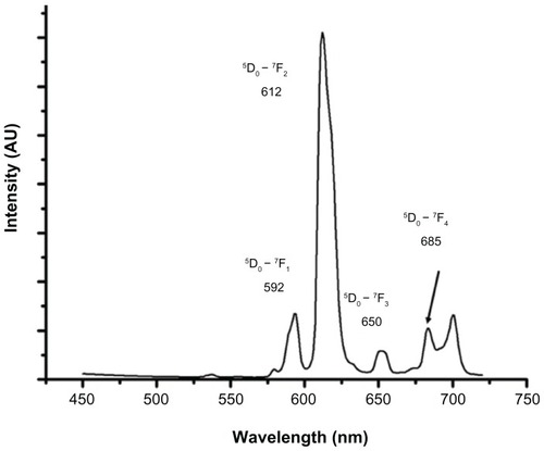
Figure 2 WST-1 proliferation assay to assess cytotoxicity of Eu-SWCNT treatment (0.2 μg/mL) for (A) SK-BR-3, (B) MCF-7, and (C) NIH 3T3 cells.
Notes: Data are presented as mean ± standard deviation (n = 3; P < 0.05 at 12-hour time point). No significant change was observed between Eu-SWCNT-treated and untreated cells at each time point.
Abbreviations: Eu-SWCNT, europium–catalyzed single–walled carbon nanotubes; MCF, Michigan Cancer Foundation breast cancer cells; WST–1, tetrazolium salt 2-(4-iodophenyl)-3-(4-nitophenyl)-5-(2,4-disulfophenyl)-2H-tetrazoilium.
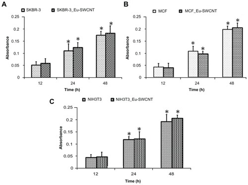
Figure 3 Confocal luminescence images of SK-BR-3 cells treated with Eu-SWCNT (0.2 μg/mL) for 24 hours at 37°C (excitation wavelength = 365 nm, emission wavelength = 560 nm, with long pass filter).
Notes: (A) Confocal images of untreated cells in (I) bright field, (II) luminescence, and (III) overlay of bright field and luminescence; (B) images of Eu-SWCNT in SK-BR-3 cells in bright field; (C) optical and luminescence images of Eu-SWCNT-internalized cells – circled in panel BI – at 40× magnification. Arrows point to a few representative Eu-SWCNTs.
Abbreviation: Eu-SWCNT, europium–catalyzed single–walled carbon nanotubes.
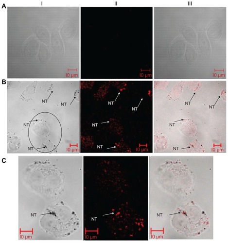
Figure 4 Confocal red luminescence images of SK-BR-3 cells treated with Eu-SWCNTs (0.2 μg/mL) for 24 hours at 37°C (excitation = 458 nm, emission = 560 nm, with long pass filter), and of untreated controls.
Notes: (A) Confocal images of Eu-SWCNT-untreated cells in the (I) bright field, (II) red luminescence, (III) nucleus stained with DAPI, and (IV) overlay of bright field and luminescence; (B) confocal images of Eu-SWCNT-treated SK-BR-3 cells in (I) bright field, (II) red luminescence, (III) nucleus stained with DAPI, (IV) overlay of Eu-SWCNT luminescence and DAPI, and (V) overlay of bright field, Eu-SWCNT luminescence, and DAPI; (C) optical and luminescence images of Eu-SWCNT-internalized and DAPI-stained cells – circled in panel BI – at 40× magnification.
Abbreviations: DAPI, diamidino-2-phenylindole; Eu-SWCNT, europium–catalyzed single–walled carbon nanotubes.
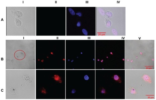
Figure 5 TEM images of Eu-SWCNT distributed in the cytoplasm and nucleus of SK-BR-3 cancer cells.
Notes: TEM images of morphology of (A) untreated cells; (B) Eu-SWCNT-treated cells; distribution in the nucleus of Eu-SWCNT at (B) low magnification and (C) high magnification; (D) presence of Eu-SWCNT in cytoplasmic vacuoles; Eu-SWCNT-treated cells, showing their distribution in (E) lysosomes (yellow circles) and (F) endosomes (purple circles). Arrows point to Eu-SWCNT.
Abbreviations: TEM, transmission electron microscopy; N, nucleus; NM, nuclear membrane; Cy, cytoplasm; V, vacuole; M, mitochondria; ST-N, Eu-SWNT in nucleus; NT-E, endocytosed Eu-SWNT.
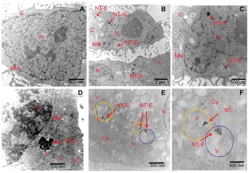
Figure 6 Raman spectra and G-band intensity map of Eu-SWCNT in SK-BR-3 cells.
Notes: (A) bright field image of the cells treated with 0.2 μg/mL of Eu-SWCNT for 24 hours. (B) Within SK-BR-3 cells, Raman G-band intensity was acquired at different points (marked in red) to detect and confirm the presence Eu-SWCNT. (C) Raman spectra of Eu-SWCNT uptaken by the cells. (D) Area map of Raman G-band intensity of Eu-SWCNT in SK-BR-3 cells. (E) Area map of Raman G-band intensity of Eu-SWCNT in cells, overlaid on an optical micrograph of the same region.
Abbreviation: Eu-SWCNT, europium–catalyzed single–walled carbon nanotubes.
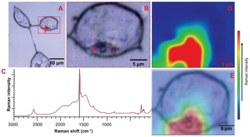
Figure 7 Cellular endocytic mechanism of Eu-SWCNT uptake: confocal fluorescence images of different cells after incubation with Eu-SWCNT at 4°C for 1 hour.
Notes: (I) Optical images; (II) Eu-SWCNT luminescence images; (III) DAPI-labeled images; (IV) overlay of Eu-SWCNT luminescence and DAPI fluorescence; (V) overlay of Eu-SWCNT luminescence, DAPI fluorescence, and optical image.
Abbreviations: DAPI, diamidino-2-phenylindole; Eu-SWCNT, europium–catalyzed single–walled carbon nanotubes; MCF, Michigan Cancer Foundation breast cancer cells; SKBR, Sloan–Kettering breast cancer cells.
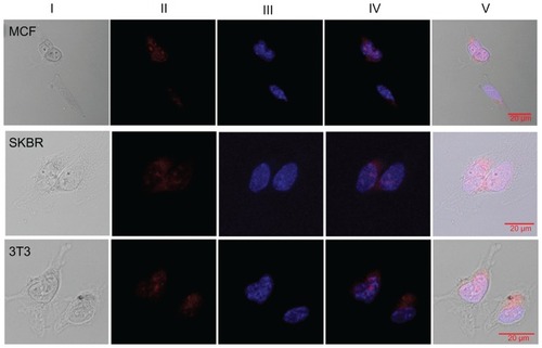
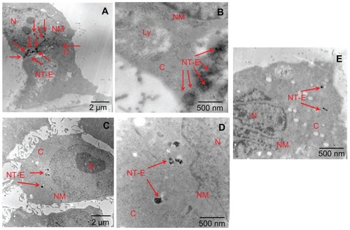
Figure 8 Transmission electron microscopy images of Eu-SWCNT-internalized SK-BR-3 cancer cells at (A and B) 37°C, (C and D) 4°C, and (E) ATP depletion conditions.
Notes: (A) Cells treated with Eu-SWCNT at 37°C showing endocytosed Eu-SWCNT distributed in the cytoplasm; (B) higher magnification showing the presence of Eu-SWCNT (red arrows); (C) low magnification, and (D) high magnification of cells maintained at 4°C, showing lesser amounts of internalized Eu-SWCNT; (E) ATP depletion conditions show lower internalization of Eu-SWCNT.
Abbreviations: N, nucleus; NM, nuclear membrane; Cy, cytoplasm; V, vacuole; M, mitochondria; ST-N, Eu-SWNT in nucleus; NT-E, endocytosed Eu-SWNT.