Figures & data
Figure 1 Structural characterization of DA-Chit. (A) Fourier transform infrared spectrum and (B) 1H nuclear magnetic resonance spectrum.
Abbreviation: DA-Chit, deoxycholic acid-modified chitosan.
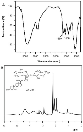
Figure 2 Water solubility of TAA and zeta potential of TAA/DA-Chit nanoparticles at various TAA-loading capacities (n = 3, mean ± standard deviation).
Abbreviations: TAA, triamcinolone acetonide acetate; DA-Chit, deoxycholic acid-modified chitosan.
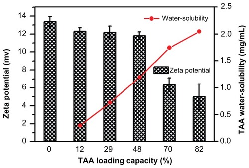
Figure 3 Transmission electron microscopy images of (A) deoxycholic acid-modified chitosan nanoparticles and triamcinolone acetonide acetate/deoxycholic acid-modified chitosan nanoparticles with different triamcinolone acetonide acetate-loading capacities, ie, (B) 12%, (C) 29%, and (D) 82%.
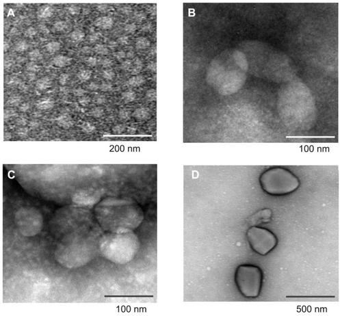
Figure 4 Morphology of cultured hRPE cells. (A) primary cultured hRPE cells, (B) Giemsa staining hRPE cells, (C) immunocytochemical staining hRPE cells, and (D) hRPE cells lacking immunohistochemical staining in control preparation (×100).
Abbreviation: hRPE, human retinal pigment epithelial.
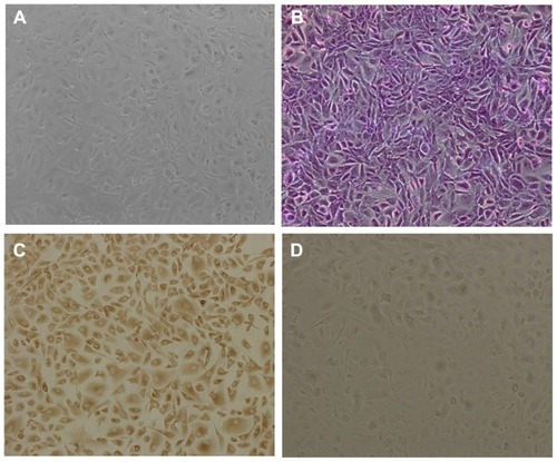
Figure 5 Cytotoxicity assay for DA-Chit at various concentrations compared with control hRPE cells (after a 24-hour incubation period, n = 5, mean ± standard deviation, *P < 0.05, statistically significant difference versus controls).
Abbreviations: DA-Chit, deoxycholic acid-modified chitosan; hRPE, human retinal pigment epithelial.
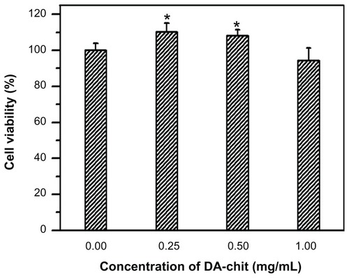
Figure 6 Cytotoxicity assay for DA-Chit, TAA, and TAA/DA-Chit nanoparticles compared with controls in human retinal pigment epithelial cells (at different incubation times and TAA concentrations of 0.1 mg/mL for TAA and TAA/DA-Chit nanoparticles, ▲P < 0.05, statistically significant difference versus controls, *P < 0.05, statistically significant difference versus the TAA group, n = 5, mean ± standard deviation).
Abbreviations: TAA, triamcinolone acetonide acetate; DA-Chit, deoxycholic acid-modified chitosan.
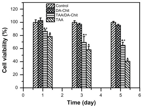
Figure 7 Typical fluorescence images of (I) hRPE cells incubated with FITC/DA-Chit nanoparticles for 24 hours at concentrations of (A) 0.3 mg/mL, (B) 0.5 mg/mL, and (C) 1.0 mg/mL; (II) hRPE cells incubated with FITC/DA-Chit nanoparticles at 1.0 mg/mL for (A) 3 hours, (B) 6 hours, and (C) 24 hours. (III) hRPE cells further incubated for different times following removal of FITC/DA-Chit nanoparticles from the culture medium, ie, (A) one day, (B) 3 days, and (C) 5 days, after incubation with 1.0 mg/mL of FITC/DA-Chit nanoparticles for 24 hours (scale bar 50 μm, 1 fluorescence field images, 2 bright field images).
Abbreviations: hRPE, human retinal pigment epithelial; FITC/DA-Chit, fluorescein isothiocyanate-labeled deoxycholic acid-modified chitosan.
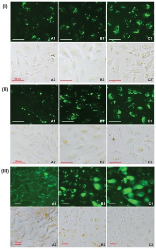
Figure 8 Cellular uptake efficiency in response to FITC/DA-Chit nanoparticles by flow cytometry analysis. (A) hRPE cells incubated with FITC/DA-Chit nanoparticles for 24 hours at concentrations of (a) 0.3 mg/mL, (b) 0.5 mg/mL, and (c) 1.0 mg/mL. (B) hRPE cells incubated with FITC/DA-Chit nanoparticles at 1.0 mg/mL for (a) 3 hours, (b) 6 hours, and (c) 12 hours. (C) Proportions of hRPE cells taking up FITC/DA-Chit versus nanoparticle concentrations. (D) Proportions of hRPE cells taking up FITC/DA-Chit nanoparticles versus incubation time.
Abbreviations: hRPE, human retinal pigment epithelial; FITC/DA-Chit, fluorescein isothiocyanate-labeled deoxycholic acid-modified chitosan; SSC, side scatter.
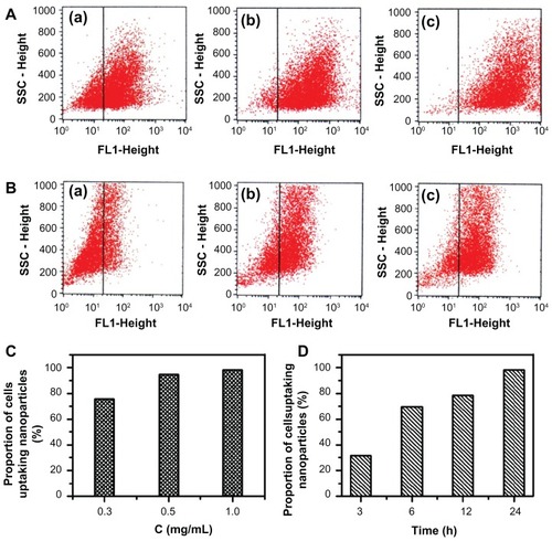
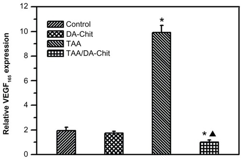
Figure 9 VEGF165 expression in hRPE cells assessed by real-time reverse transcription polymerase chain reaction (*P < 0.01, statistically significant difference versus controls; ▲P < 0.05, statistically significant difference versus the TAA-treated group).
Abbreviations: hRPE, human retinal pigment epithelial; TAA, triamcinolone acetonide acetate; DA-Chit, deoxycholic acid-modified chitosan; VEGF, vascular endothelial growth factor.