Figures & data
Figure 1 High-resolution transmission electron microscopic image of an isolated zerovalent bismuth nanoparticle.
Notes: The colloidal sample to obtain this image was prepared a few minutes before the microscopy session. Final concentrations of the chemical reagents are 2 × 10⁻³ M Bi(NO3)3 · 5H2O, 4 × 10⁻⁴ M Na3(C6H5O7) · 2H2O, and 4 × 10⁻⁴ M NaBH4 in dimethyl sulfoxide.
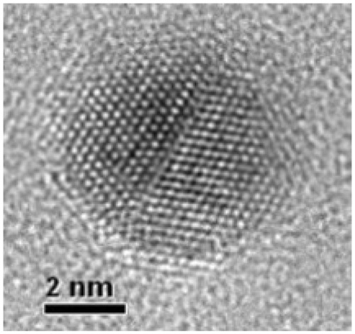
Figure 2 Antimicrobial activity of zerovalent bismuth nanoparticles against Streptococcus mutans growth.
Notes: The y axis shows the optical density units of S. mutans growth. S. mutans culture without any inhibitor was used as growing control and chlorhexidine 0.12% as a positive inhibition control. Zerovalent bismuth nanoparticles were used at a final concentration of 2 mM.
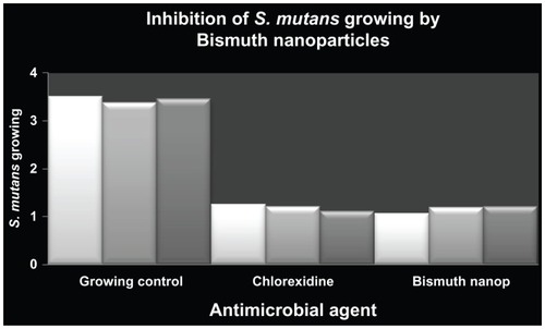
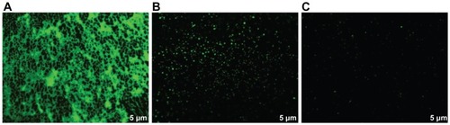
Figure 3 Inhibition of Streptococcus mutans biofilm detected by fluorescence microscopy.
Notes: S. mutans culture without any inhibitor was used as growing control and chlorhexidine 0.12% as a positive inhibition control. Zerovalent bismuth nanoparticles were used at a final concentration of 2 mM.