Figures & data
Figure 1 Classification of surfactants and structures of the ionic and nonionic surfactants mentioned in this review.
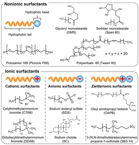
Figure 2 (A) Typical Illustration of surfactant-coated nanoparticles. (B) Various organic and inorganic materials used in the core of surfactant-coated nanoparticles.
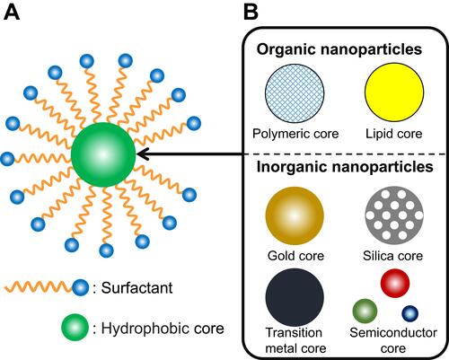
Figure 3 (A) Equations of the DLVO theory. (B) Relationship between two particles assuming the DLVO theory. (C) A typical example of potential energy presented in the DLVO theory.
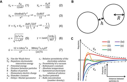
Table 1 Summary of Surfactant-Coated Nanoparticles Used in the Field of Nanomedicine
Figure 4 Behavior and fate of surfactant-coated nanoparticles in the blood stream.
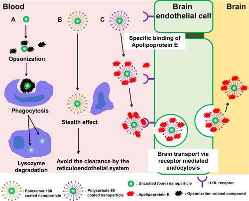
Figure 5 Active and passive targeting of nanoparticles to the cancer cells.
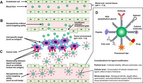
Table 2 Summary of Surfactant-Coated Nanoparticles Used in the Field of Food Nanotechnology
Figure 6 The potential applications of food nanotechnology.
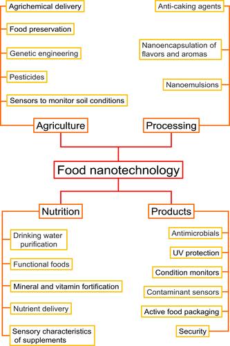
Figure 7 Illustration of the relationships between diseases, free radicals, reactive oxygen species, and aging in the body, and its regulation by antioxidants from food source.
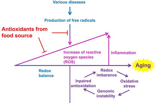
Figure 8 Age-related decrease in plasma concentrations of antioxidants from food source.
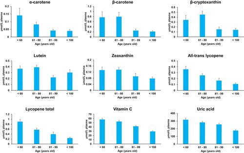
Figure 9 The digestive stages after oral administration and the mechanisms of in vivo uptake of surfactant-coated nanoparticles through the small intestine.
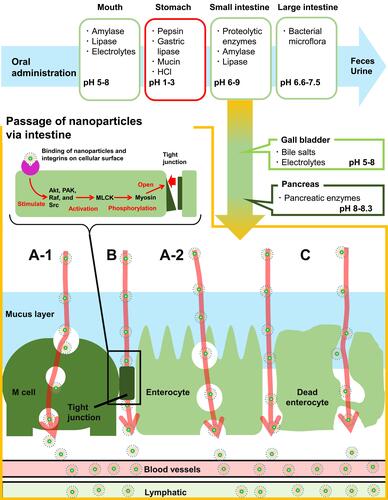
Figure 10 A wide variety of nanoscale materials potentially present in foods from both natural and artificial sources.
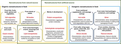
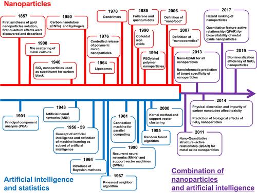
Figure 11 Overlapping timelines of the development of artificial intelligence and nanomaterials. Since 2010, these two fields have developed a powerful synergy.