Figures & data
Figure 1 The chemical structures of (A) celastrol and (B) PEG-b-PCL nanopolymeric micelles. (C) In vitro drug release profiles of celastrol from PEG-b-PCL micelles (celastrol content: 7.36%) in phosphate buffer solutions (pH 7.4) and sodium acetate buffered solution (pH 5.0) at 37°C.
Notes: A typical two-phase-release profile contained a rapid release in the first stage followed by a sustained and slow release over a prolonged time up to several weeks in both solutions. Furthermore, celastrol release at pH 5.0 was significantly faster than that at pH 7.4. The data are presented as the means ± standard deviation (n = 3).
Abbreviation: PEG-b-PCL, poly(ethylene glycol)-block-poly(ɛ-caprolactone).
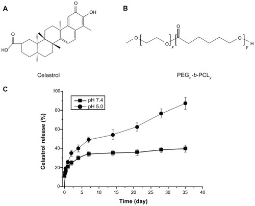
Figure 2 CNPs inhibit the proliferation of human SO-Rb 50 cells in vitro. (A) Morphological variations of SO-Rb 50 cells treated with 0–54.4 μg/mL CNPs for 48 hours (100 × magnification). Increasing the amount of CNPs decreased the number and size of SO-RB 50 cells. (B) The dose-dependent effect of CNPs on cell viability. SO-Rb 50 cells were treated with 0–54.4 μg/mL CNPs for 48 hours. The IC50 is 17.733 μg/mL when exposed to CNPs for 48 hours. (C) The time-dependent effect of CNPs on the cell viability. SO-Rb 50 cells were, respectively, treated with 27.2 μg/mL CNPs and 2 μg/mL free celastrol dissolved in 0.2% (v/v) DMSO for 0, 24, 48, and 72 hours.
Notes: The medium (0.2% DMSO) and blank micelles without celastrol showed a cell viability of greater than 95%. The results are reported as the means ± standard deviation (n = 3) which were performed in triplicate. Statistical significance was based on the difference compared to control cells treated with medium (*P < 0.01, error bar, 95% CI).
Abbreviations: CI, confidence interval; CNPs, celastrol nanoparticles; Cel, celastrol; DMSO, dimethyl sulfoxide.
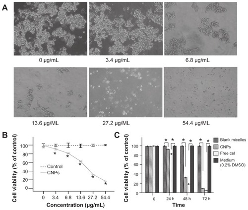
Figure 3 CNPs induce apoptosis in SO-Rb 50 cells. Fluorescence micrographs of SO-Rb 50 cells are shown after exposure to (A) blank micelles and (B) CNPs at 54.4 μg/mL for 24 hours (400× magnification).
Notes: Cells were stained with Hoechst 33342 to visualize nuclear morphology. Blank micelle-treated SO-Rb 50 cells have round nuclei with homogeneous chromatin. The cells treated with CNPs are characterized by chromatin condensation, a reduction of nuclear size, and nuclear fragmentation (red arrows show nuclear fragmentation).
Abbreviation: CNPs, celastrol nanoparticles.
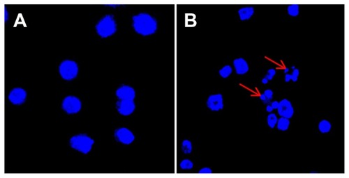
Figure 4 Annexin V-FITC/PI double staining analysis of apoptosis in SO-Rb 50 cells at 24 hours post-treatment. (A) Dot plot for flow cytometric analysis of apoptotic cells. Living cells tested negative for both annexin V-FITC and PI. Populations testing annexin V positive/PI negative were classified as early-stage apoptotic cells, and double-positive cells were classified as late-stage apoptotic cells. (B) Bar graph quantifying the percentage of dead, living, early-stage apoptotic, and late-stage apoptotic cells according to treatment.
Note: The values shown are the mean of three independent experiments.
Abbreviations: FITC, fluorescein isothiocyanate; PI, propidium iodide; CNPs, celastrol nanoparticles.
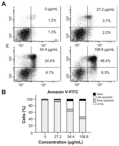
Figure 5 Effect of CNPs on the expression level of Bcl-2, Bax, NF-κB p65, and phospho-NF-κB p65. The SO-Rb 50 cells were incubated with blank micelles or CNPs (54.4 μg/mL) for 48 hours. Western blot analyses were performed with antibodies against Bcl-2, Bax, NF-κB p65, and phospho-NF-κB p65.
Notes: GAPDH expression is shown as a loading control. This figure is representative of three independent experiments. The data are presented as means ± standard deviation. Statistical significance is based on the difference when compared with blank micelle-treated cells (*P < 0.001).
Abbreviations: CNPs, celastrol nanoparticles; GAPDH, glyceraldehyde 3-phosphate dehydrogenase.
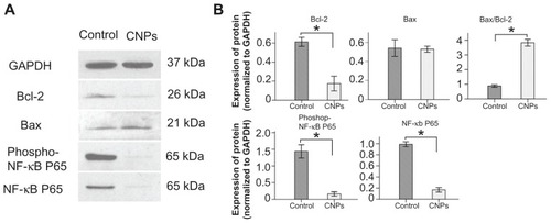
Figure 6 CNPs suppress retinoblastoma growth in a xenograft mouse model. SO-Rb 50 cells were subcutaneously injected into 6-week-old NOD SCID mice (5 × 10⁷ per mouse). The mice were administrated CNPs or blank micelles (27.2 mg/kg/2 days) by intraperitoneal injections after solid tumors grew to 100 mm3. (A) CNPs inhibit tumor growth as measured by tumor volume. (B) As demonstrated by the change in body weight, CNPs had little toxicity in mice at the doses tested. (C) Solid tumors in mice treated with CNPs were significantly smaller than those in control mice.
Notes: *P < 0.001 versus control. Error bars: 95% CI.
Abbreviation: CNPs, celastrol nanoparticles.
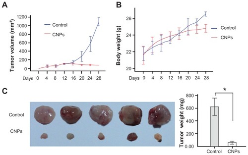
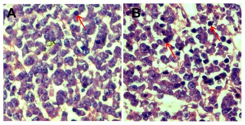
Figure 7 CNPs induce apoptosis in retinoblastoma cells. Retinoblastoma induced by inoculation with SO-Rb 50 cells were treated with blank micelles and CNPs at 27.2 mg/kg every other day for 3 weeks. Retinoblastoma tissue sections of (A) control and (B) CNP groups were stained with hematoxylin and eosin.
Note: A greater number of cells showed a reduction in nuclear size, deeply stained nuclei, and condensed cytoplasm in the CNP group (red arrows show hyperchromatic nuclei).
Abbreviation: CNPs, celastrol nanoparticles.