Figures & data
Figure 1 Illustration of mechanism of (A) MRI and (B) SSB examination for antibody-mediated magnetic nanoparticles on liver tumor tissue.
Abbreviations: AC, alternating current; MRI, magnetic resonance imaging; SSB, scanning superconducting-quantum-interference-device biosusceptometry.
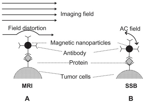
Table 1 Information of in-vivo tests and biopsy tests in this work
Figure 2 Characteristics of anti-CEA MF, anti-AFP MF, and MF without antibody coatings: (A) the hydrodynamic diameters, (B) saturation magnetism, (C) the stability of antibody-mediated MFs.
Abbreviations: CEA, carcinoembryonic-antigen; MF, magnetic fluid; AFP, alphafetaprotein.
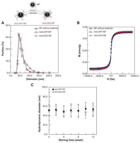
Figure 3 MRI examination of a liver tumor. (A) Photos of the test mice and representative MRI images of the liver tumor at different examination time. (B) The variation ratio of the average normalized intensity ΔI/I0 and the variation of image contrast ΔC/C0.
Abbreviations: AFP, alphafetaprotein; CEA, carcinoembryonic-antigen; MF, magnetic fluid; MRI, magnetic resonance imaging.
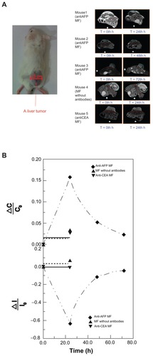
Figure 4 SSB examination of a liver tumor. (A) Setup scheme. (B) The scanning curves of all test mice at different times. (C) The variation of magnetism M of all test mice at different times. (D) The analysis comparison of SSB and MRI.
Abbreviations: AFP, alphafetaprotein; CEA, carcinoembryonic-antigen; MF, magnetic fluid; SSB, scanning superconducting-quantum-interference-device biosusceptometry; MRI, magnetic resonance imaging.
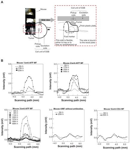
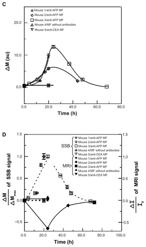
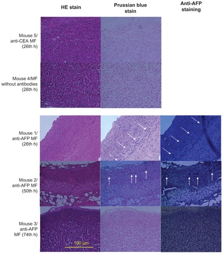
Figure 5 HE stain and Prussian blue stain of liver tumor tissue after the injection of MFs (magnification ×400).
Abbreviations: HE, hematoxylin and eosin; AFP, alphafetaprotein; MF, magnetic fluid; CEA, carcinoembryonic antigen.