Figures & data
Figure 1 (A) Schematic illustration of nanoparticle formation, and (B) transmission electron microscopy image of dextran-spermine-plasmid DNA nanoparticles.
Note: The concentration of plasmid DNA is 10 μg/mL.
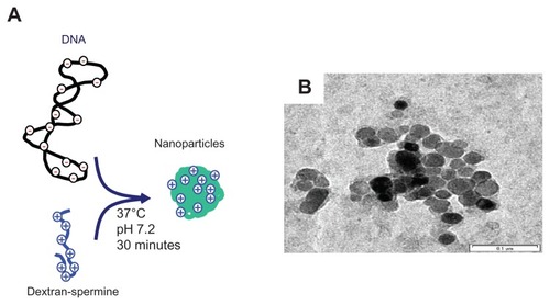
Table 1 Characterization of plasmid DNA, dextran-spermine, and DNA nanoparticles
Figure 2 In vitro release profiles of plasmid DNA-BMP-2 from DNA nanoparticles incorporated into nanofibers (●). Naked plasmid DNA-BMP-2 incorporated into nanofibers (▴) was used as the control group.
Note: †P < 0.05; significant against the amount of plasmid DNA released from naked plasmid DNA incorporated into nanofibers.
Abbreviation: BMP-2, bone morphogenic protein-2.
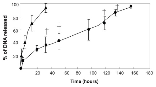
Figure 3 Proliferation of mesenchymal stem cells on three-dimensional collagen-PGA nanofiber sheet (■) and two-dimensional tissue culture plate (●).
Note: †P < 0.05; significant relative to the two-dimensional culture system.
Abbreviation: PGA, poly (glycolic acid).
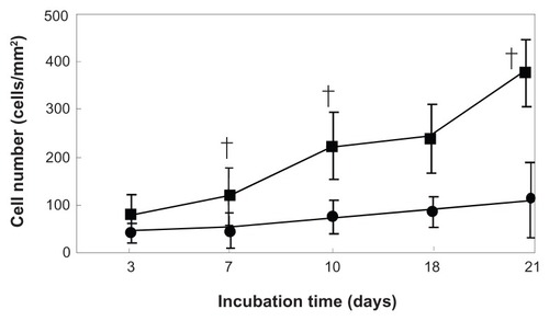
Table 2 BMP-2 activity of MSC 2 days after treatment of free plasmid DNA and nanoparticles of dextran-spermine-plasmid DNA
Figure 4 (A) Schematic illustration of electrospinning machine. (B) Cross-sectional scanning electron microscopy photographs of the structural morphology of PGA/collagen nanofibers fabricated by electrospinning.
Abbreviation: PGA, poly (glycolic acid).
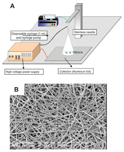
Figure 5 Cross-sectional scanning electron microscopy photographs of the structural morphology of PGA/collagen nanofibers fabricated by electrospinning (A) 1 week, (B) 2 weeks, and (C) 3 weeks after culturing the mesenchymal stem cells on nanofibers incorporated with dextran-spermine-plasmid DNA nanoparticles.
Abbreviation: PGA, poly (glycolic acid).
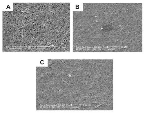
Table 3 Biochemical analysis of the osteogenic differentiation of genetically engineered mesenchymal stem cells