Figures & data
Figure 1 Schematic presentation of various subtypes of extracellular vesicles such as exosomes, ectosomes and apoptotic bodies, are released into the extracellular environment during physiological and pathological processes.
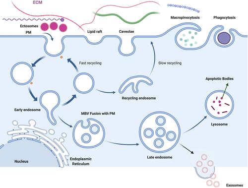
Figure 2 Biogenesis of exosomes by ESCRT dependent mechanism and independent mechanism involved with accessory proteins and lipid dependent pathway.
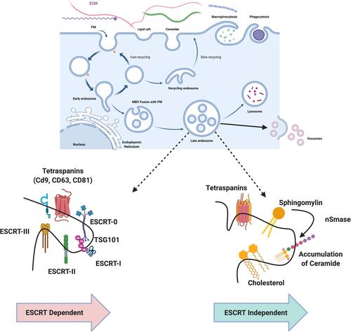
Figure 3 Mechanism of biogenesis of ectosomes. Increased level of accumulation of Ca2+ at the plasma membrane and involvement of translocase enzymes and proteins such as ADP-ribosylation factor 6 (ARF6), extracellular signal regulated kinases (ERK) and phosphorylation of myosin light chain kinase (MLCK). (A) Extensive accumulation of Ca2+ at the PM region causes the imbalance of the phospholipids orientation. (B) Role of flippase and floppase to maintains the phospholipids symmetry. (C) ARF6 activates ERK followed by the phosphorylation of myosin light chain which stimulates the budding of ectosomes from the PM.
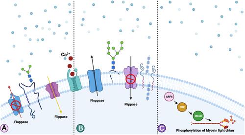
Figure 4 Sorting of cargoes into EVs. Proteins and RNA can be packaged into the EVs by various mechanisms including ubiquitination, phosphorylation, myristoylation, glycosylation and sumoylation.
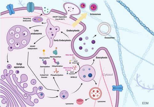
Figure 5 Schematic representation of isolation of EVs from conditioned medium using various centrifugation steps and isolated EVs displayed packaging of cargoes DNA, proteins, miRNA, antibodies, tetraspanins, histone, actin and tubulin.
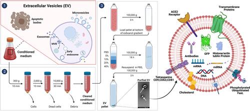
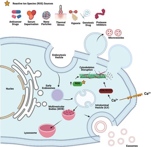
Figure 6 Various sources of oxidative agents responsible for oxidative stress such as anticancer drug, serum deprivation, nanoparticles, thermal stress, hypoxia, genotoxic drugs and protease inhibitors induce biogenesis and release of EVs.