Figures & data
Figure 1 X-ray patterns of the CONPs.
Note: Results revealed the crystal structure of the CONPs, and no characteristic peaks of impurities were detected.
Abbreviation: CONPs, cuprous oxide nanoparticles.
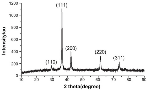
Figure 2 UV-vis spectra of the CONPs.
Note: CONPs were dissolved in deionized water and deionized water was used as a reference.
Abbreviation: CONPs, cuprous oxide nanoparticles.
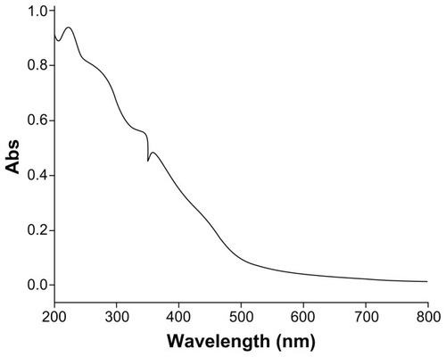
Figure 3 FTIR transmission spectra of the CONPs. The band at 624 cm−1 correlates to the stretching vibration of the CONPs.
Notes: Moreover, there were also some stretching bands. The other bands at 3398 cm−1, 2992 cm−1, 1373 cm−1, and 839 cm−1 are probably due to the carbonate moieties and water that are generally observed, when FTIR samples are measured in air.
Abbreviations: CONPs, cuprous oxide nanoparticles; FTIR, Fourier transform-infrared.
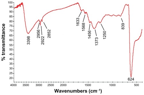
Figure 4 TEM images of the CONPs.
Note: The CONPs were all perfect spheres, and the mixtures were homogeneous.
Abbreviations: CONPs, cuprous oxide nanoparticles; TEM, transmission electron microscopy.
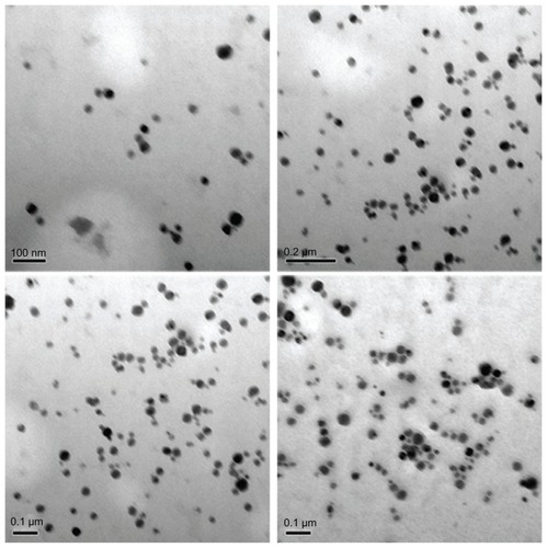
Figure 5 Differential cytotoxicity of CONPs on tumor cell (melanoma cells YUMAC), paired control tumor cell (HeLa), and normal cells (293T and MEF) detected by MTT assay.
Notes: HeLa cells and melanoma cells were more sensitive, and the two normal cell lines (293T and MEF cells) were tolerant, especially at low concentrations. Cells were treated with the CONPs for 48 hours (*P < 0.005, n = 3).
Abbreviations: CONPs, cuprous oxide nanoparticles; MEF, mouse embryonic fibroblast.
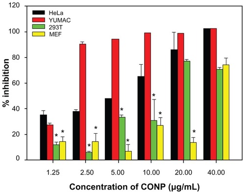
Figure 6 Flow cytometry analysis of HeLa and MEF cells treated with different concentrations of CONPs for 48 hours and then stained with Annexin V and PI.
Notes: Significant increases in apoptosis were observed in HeLa cells exposed to the 10 μg/mL and 20 μg/mL CONPs for 48 hours. However, decreased apoptosis was observed in the MEF cells. The upper-left quadrant in each panel shows the necrotic population, the upper-right quadrant shows the late apoptotic population, and the lower-right quadrant shows the early apoptotic population.
Abbreviations: CONPs, cuprous oxide nanoparticles; MEF, mouse embryonic fibroblast; PI, propidium iodine.
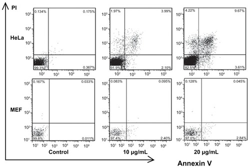
Figure 7 Cell cycle progression assay of HeLa cells treated with different concentrations of CONPs for 48 hours.
Notes: Proliferation of HeLa cells was strongly suppressed by CONPs in a dose-dependent manner. Cells were arrested in the G1/G0 phase after 48 hours of treatment and were less able to proliferate.
Abbreviation: CONPs, cuprous oxide nanoparticles.
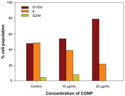
Figure 8 Transmission electron images of HeLa cells treated with a solution of CONPs (30 μg/mL) for 4 hours. The arrows indicate the accumulation of the CONPs. (A) The CONPs entered a mitochondrion and broke its outer membrane. CONPs existed in vesicles and in the cytoplasm. (B) The endoplasmic reticulum swelled visibly. A structure similar to an autophagy body phagocytized a group of CONPs. (C) The vesicles containing the CONPs appeared to target the outer membrane of the mitochondrion, and the shape of the mitochondrion had changed. (D) CONPs localized in small vesicles and entered or were entering the mitochondrion. (E and F) CONPs broke the membrane of the mitochondrion and caused structural changes.
Abbreviations: CONPs, cuprous oxide nanoparticles; M, mitochondrion; ER, endoplasmic reticulum.
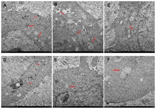
Figure 9 DTT protection experiment on HeLa cells for 48 hours. The MTT assay was then performed to evaluate protective effects of DTT.
Notes: The results show that DTT could increase cell viability at low concentrations but reduced viability at higher concentrations (P < 0.01, n = 3).
Abbreviations: CONPs, cuprous oxide nanoparticles; DTT, dithiothreitol; MTT, 3-(4,5-di-methylthiazol-2yl)-2,5-diphenyl tetrazolium bromide.
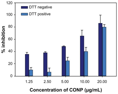
Figure 10 Lipid peroxidation measurement of CONPs in the liposomes prepared from soybean lecithin and cholesterol.
Notes: CONPs increased the lipid peroxidation activity and produced ROS, but DTT could significantly block the generation of ROS (P < 0.01, n = 3).
Abbreviations: CONPs, cuprous oxide nanoparticles; DTT, dithiothreitol; ROS, reactive oxygen species.
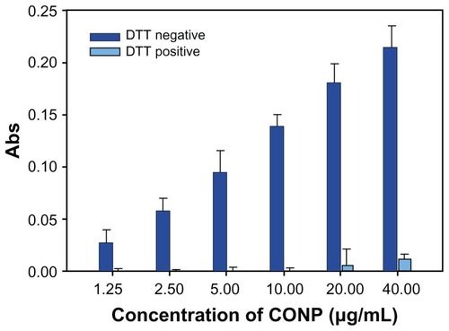
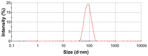
Figure S1 Hydrodynamic size distributions of CONPs in water.
Note: The diameter of the CONPs ranged from approximately 40 nm to 110 nm.
Abbreviation: CONPs, cuprous oxide nanoparticles.