Figures & data
Table 1 The Independent Variables Levels Used to Formulate CLT Loaded SPs Utilizing (32) Complete Factorial Design
Table 2 Measured Responses of CLT Formulations of the Experimental Complete 32 Factorial Design
Table 3 The Statistics Summary of Complete Factorial Design (32) Used for Optimization of SPs Formulations
Figure 1 Linear correlation plots presenting the effect of (A) Span 60: EA ratio and (B) EA type on the following parameters: EE%, VS, PDI and ZP.
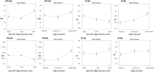
Figure 2 The optimum SPs formulation (S1) transmission electron micrograph.
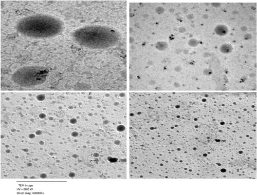
Table 4 Effect of Storage on Physical Properties of the Optimum Formulation S1
Figure 3 In vitro release study of CLT formulations.
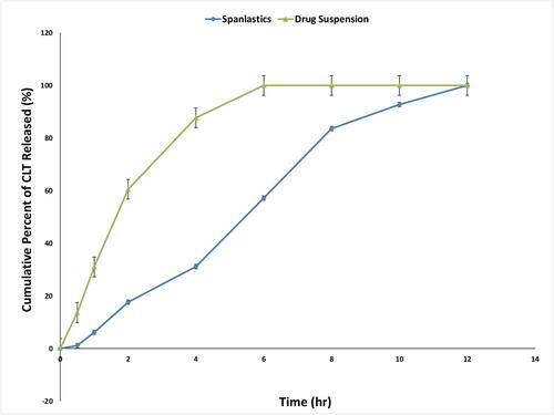
Table 5 Kinetics of CLT Release from S1 According to Different Kinetic Models
Figure 4 Ex vivo corneal permeability of CLT formulations.
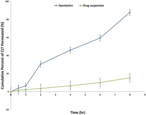
Figure 5 Microbiological assay of (A) CLT suspension, (B) Non-medicated S1 and (C) S1 for the treatment of Candida albicans infection. Data are presented as mean ± SD, (n=3).
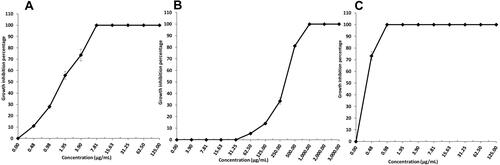
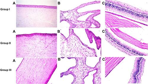
Figure 6 Photomicrographs presenting histopathological sections (stained by hematoxylin and eosin) of normal untreated rabbit eye (group 1), rabbit eye treated with CLT suspension (group 2) and rabbit eye treated with S1 (group 3). (A) Shows histological structure of the cornea, (B) Shows histological structure of the iris and (C) Shows histological structure of the sclera, retina and choroid.