Figures & data
Figure 1 Mean diameters of nanoparticles of mPEG45-PH15-PLLA82 (A) and mPEG45-PH30-PLLA82 (B), and zeta potential of mPEG45-PH15-PLLA82 (C) and mPEG45-PH30-PLLA82 (D) nanoparticles at different pH values.
Abbreviation: mPEG-PH-PLLA, methoxyl poly(ethylene glycol)-poly(L-histidine)-poly(L-lactide).
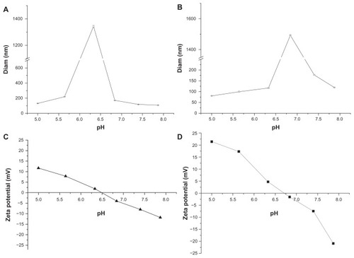
Figure 2 Transmission electron microscopic images of nanoparticles at different pH levels. (A) and (B) are mPEG45-PH15-PLLA82 nanoparticles. (C) and (D) are mPEG45-PH30-PLLA82 nanoparticles. The pH value of (A) and (C) was 5.0 and that of (B) and (D) was 7.4.
Abbreviation: mPEG-PH-PLLA, methoxyl poly(ethylene glycol)-poly(L-histidine)- poly(L-lactide).
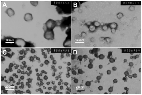
Figure 3 Release profiles of doxorubicin-loaded mPEG45-PH15-PLLA82 and mPEG45- PH30-PLLA82 nanoparticles at different pH values.
Abbreviations: DOX, doxorubicin; mPEG-PH-PLLA, methoxyl poly(ethylene glycol)-poly(L-histidine)-poly(L-lactide).
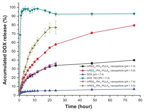
Figure 4 Cell viability of mPEG45-PH15-PLLA82 and mPEG45-PH30-PLLA82 nanoparticles incubated with NIH 3T3 fibroblasts for 24 hours.
Abbreviation: mPEG-PH-PLLA, methoxyl poly(ethylene glycol)-poly(L-histidine)- poly(L-lactide).
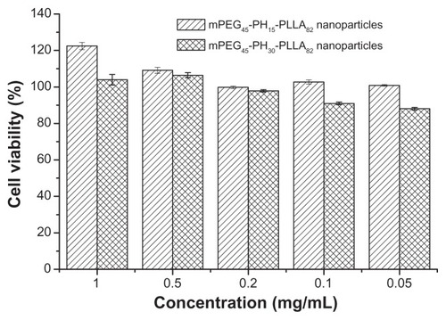
Figure 5 Cell viability of mPEG45-PH15-PLLA82 and mPEG45-PH30-PLLA82 nanoparticles incubated with HepG2 cells for 24 hours.
Abbreviation: mPEG-PH-PLLA, methoxyl poly(ethylene glycol)-poly(L-histidine)- poly(L-lactide).
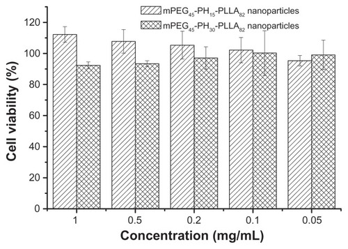
Figure 6 Confocal microscopy photographs of HepG2 cells incubated with doxorubicin-loaded nanoparticles. (A1–A4) doxorubicin for 0.5 hours; (B1–B4) doxorubicin-loaded mPEG45-PH15-PLLA82 nanoparticles for 0.5 hours; (C1–C4) doxorubicin-loaded mPEG45-PH30-PLLA82 nanoparticles for 0.5 hours; (D1–D4) doxorubicin hydrochloride for 0.5 hours; (E1–E4) doxorubicin for 6 hours; (F1–F4) doxorubicin-loaded mPEG45-PH15-PLLA82 nanoparticles for 6 hours; (G1–G4) doxorubicin-loaded mPEG45- PH30-PLLA82 nanoparticles for 6 hours; (H1–H4) doxorubicin hydrochloride for 6 hours. Note: The four photographs from left to right are red doxorubicin, stained nucleus, bright field, and overlapped graphs.
Abbreviation: mPEG-PH-PLLA, methoxyl poly(ethylene glycol)-poly(L-histidine)- poly(L-lactide).
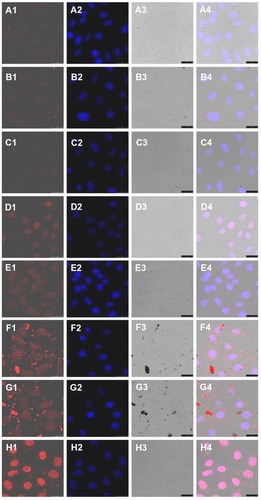
Figure 7 Flow cytometry results of doxorubicin, doxorubicin hydrochloride, and doxorubicin-loaded mPEG45-PH15-PLLA82 and mPEG45-PH30-PLLA82 nanoparticles incubated with HepG2 cells at 37°C for 6 hours (doxorubicin concentration 10 μg/mL).
Notes: 1, control; 2, doxorubicin; 3, doxorubicin hydrochloride; 4, doxorubicin-loaded mPEG45-PH15-PLLA82 nanoparticles; 5, doxorubicin-loaded mPEG45-PH30- PLLA82 nanoparticles.
Abbreviation: mPEG-PH-PLLA, methoxyl poly(ethylene glycol)-poly(L-histidine)- poly(L-lactide).
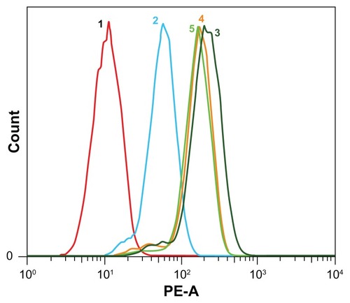
Figure 8 Morphology of HepG2 cells incubated with doxorubicin-loaded nanoparticles, the time was 36 hours and the concentration of doxorubicin was 15 μg/mL. (A) Cell culture plate, (B) doxorubicin hydrochloride, (C) doxorubicin, (D) doxorubicin-loaded mPEG45-PH15-PLLA82 nanoparticles, and (E) doxorubicin-loaded mPEG45-PH30-PLLA82 nanoparticles.
Abbreviation: mPEG-PH-PLLA, methoxyl poly(ethylene glycol)-poly(L-histidine)-poly(L-lactide).
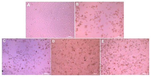
Figure 9 In vitro antitumor activity of doxorubicin-loaded nanoparticles incubated with HepG2 cells, drug concentration was 15 μg/mL.
Abbreviations: DOX, doxorubicin; mPEG-PH-PLLA, methoxyl poly(ethylene glycol)-poly(L-histidine)-poly(L-lactide).
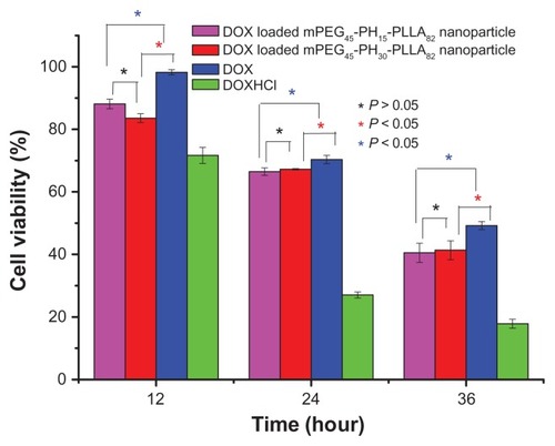
Figure 10 In vitro antitumor activity of doxorubicin-loaded nanoparticles with different drug concentrations, incubation time was 36 hours.
Abbreviations: DOX, doxorubicin; mPEG-PH-PLLA, methoxyl poly(ethylene glycol)- poly(L-histidine)-poly(L-lactide).
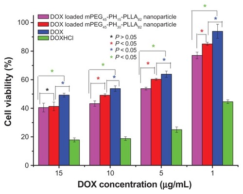
Figure S1 The synthetic route of mPEG-PH-PLLA triblock copolymer.
Abbreviations: NHS, N-Hydroxysuccinimide; DCC, N,N’-Dicyclohexylcarbodiimide; MEE, methoxyethoxyethanol.
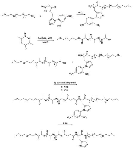
Figure S2 The 1H NMR spectra of mPEG45-NH2, mPEG45-PH30 and mPEG45-PH30-PLLA82.
Abbreviation: ppm, parts per million.
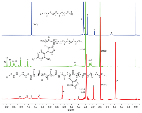
Table S1 Characterizations of mPEG-PH-PLLA triblock copolymer nanoparticles