Figures & data
Table 1 Amount of Free Thiol Groups and Disulfide Bonds Immobilized on EC–Cysteamine
Figure 1 Oxidation and reduction amination, EC (A), EC-CHO (B), EC-cysteamine (C).
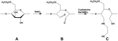
Figure 2 Mucoadhesive properties of EC, EC-CHO and EC-cysteamine; indicated values are means of at least three experiments±SD.
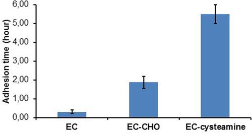
Figure 3 Viscosity values and rheological synergism for 3% (m/v) EC, EC-CHO and EC-cysteamine and their corresponding mixtures with mucin; indicated values are means of at least three experiments±SD.
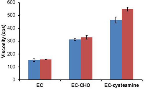
Figure 4 Glass transition (Tg) curve of EC.
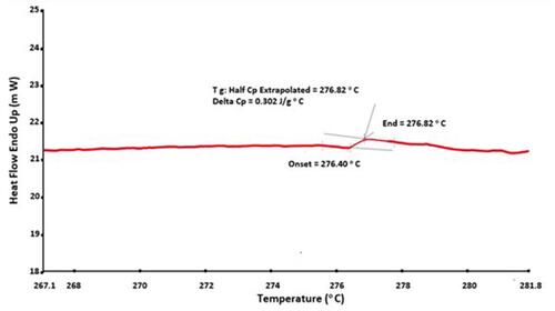
Figure 5 Glass transition (Tg) curve of EC-cysteamine.
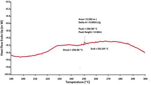
Figure 6 The morphology of EC-cysteamine nanoparticles alone (A) and EC-cysteamine nanoparticles loaded with rhodamine 123 (B).
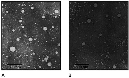
Figure 7 Degradation of EC-cysteamine (▲) and EC-cysteamine nanoparticles (X); indicated values are means of at least three experiments±SD.
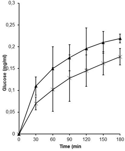
Figure 8 Cytotoxicity studies of EC, EC-cysteamine and EC-cysteamine nanoparticles using resazurin; indicated values are means of at least three experiments±SD.
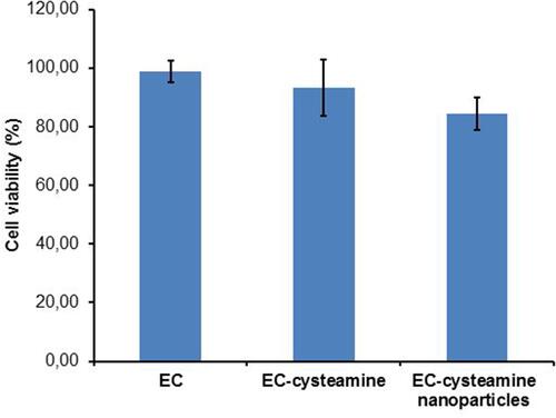
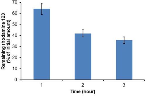
Figure 9 Release studies of EC-cysteamine nanoparticles loaded with rhodamine 123; indicated values are means of at least three experiments±SD.
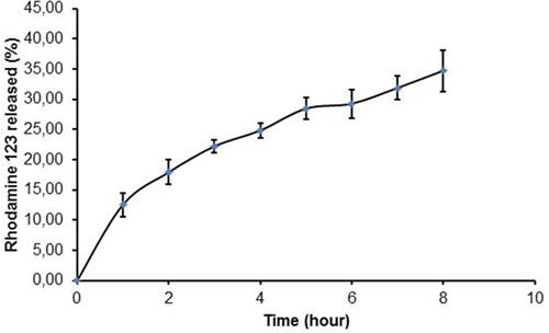
Figure 10 Mucoadhesive properties of EC-cysteamine nanoparticles; indicated values are means of at least three experiments±SD.