Figures & data
Figure 1 (A) Synthesis of MNPs and immunofunctionalized MNPs. (B) The schema illustrates the process for detection of specific T helper cells using two biomarkers: immunofunctionalized MNPs for immunoseparation and the ELISA technique for detection. Absorbance was recorded at a wavelength of 650 nm. (C) Diagrammatic representations of the T helper cells, B cells, and other materials.
Abbreviations: MNPs, magnetite nanoparticles; ELISA, enzyme-linked immunosorbent assay; HRP-stv, horseradish peroxidase-streptavidin; TMB, 3,3′,5,5′-tetramethylbenzidine.
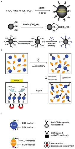
Figure 2 Typical transmission electron microscopy images of (A) synthesized MNPs and (B) APTES-modified MNPs, with (C) a bar chart indicating their respective average diameters.
Abbreviations: MNPs, magnetite nanoparticles; APTES, 3-aminopropyltriethoxysilane.
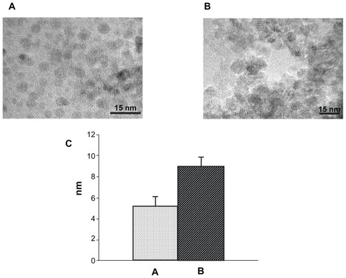
Figure 3 Synchrotron XRD spectra of (A) α-Fe2O3, (B) Fe3O4, (C) MNPs, and (D) APTES-modified MNPs.
Abbreviations: XRD, X-ray diffraction; MNPs, magnetite nanoparticles; APTES, 3-aminopropyltriethoxysilane.
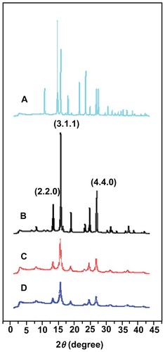
Figure 4 Synchrotron Fourier transform infrared spectra of (A) MNPs, (B) APTESmodified MNPs, and (C) immunofunctionalized MNPs.
Abbreviations: MNPs, magnetite nanoparticles; APTES, 3-aminopropyltriethoxysilane.
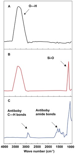
Figure 5 (A) Optical microscopic images indicating the specificity of immunofunctionalized MNPs for the T helper cells during magnetic separation and (B) a bar chart indicating the separation efficiency of immunofunctionalized MNPs at various T cell counts.
Abbreviation: MNPs, magnetite nanoparticles.
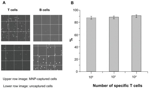
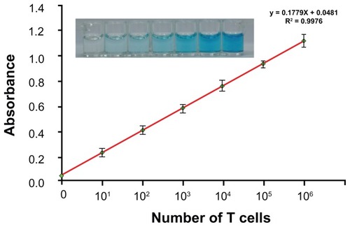
Figure 6 Semi-log plot of absorbance and the number of T helper cells detected by ELISA in a mixed cell population.
Abbreviation: ELISA, enzyme-linked immunosorbent assay.