Figures & data
Figure 1 Ceria ENM morphology. (A) Ceria ENM imaged using TEM. The insert at the top right shows the crystallinity of the ceria ENM. (B) Volume-based particle size distribution for ceria ENM of a representative batch of ceria aqueous dispersion.
Abbreviations: ENM, engineered nanomaterial; TEM, transmission electron microscopy.
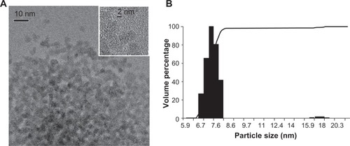
Figure 2 Ceria ENM hydrodynamic diameter in perfusate. Hydrodynamic diameter (intensity weighted average) of ceria ENM in the in situ perfusate, from 1 to 230 minutes (1000 μg/mL at 37°C) after ceria was added to the perfusate.
Notes: Solid circles are DLS data; black solid line is the average of all the data. Dotted lines represent the average diameter ± 1 SD.
Abbreviations: ENM, engineered nanomaterial; DLS, dynamic light scattering; Dmean, diameter mean.
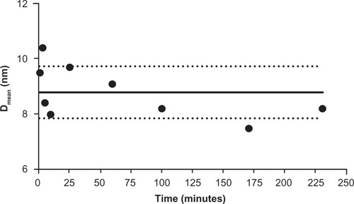
Figure 3 Vascular space of the brain as measured by Gd-DTPA at 15 (n = 4) and 20 (n = 5) mL/minute flow rate in control rats and 20 (n = 10) mL/minute flow rate in 5 nm ceria-treated rats.
Note: *Significantly different from eight brain regions, P < 0.05.
Abbreviation: Gd-DTPA, gadolinium-diethylenetriamine pentaacetic acid.
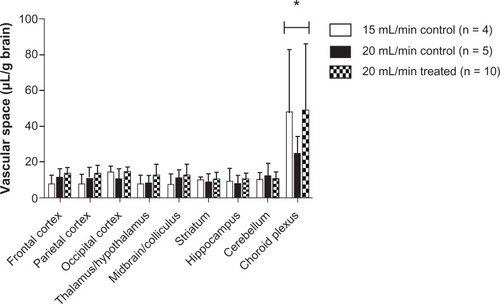
Figure 4 Ceria ENM uptake space at three concentrations and perfusion duration times. Effect of ceria ENM concentration and perfusion duration on its uptake. (A) Q uptake space (mL/g) of a 5 nm ceria ENM in eight brain regions and the choroid plexus for three concentrations, at a flow rate of 20 mL/minute, and 120-second perfusion duration. (B) Mass amount (μg/g) of 5 nm ceria ENM in eight brain regions and the choroid plexus after brain perfusion with three ceria concentrations, at a flow rate of 20 mL/minute and perfusion duration of 120 seconds. (C) Q uptake space (mL/g) of 5 nm ceria ENM in eight brain regions and choroid plexus after 20-, 60-, and 120-second perfusion at 30 μg/mL, at a flow rate of 20 mL/minute.
Notes: (A) #Significantly different among three concentration groups, P < 0.05; *significantly different compared to the eight brain regions at the same concentration, P < 0.05. (B) #Significantly different compared to 30 μg/mL concentration group; *significantly different compared to the eight brain regions, P < 0.05. (C) #Significantly different between 20- and 60-second perfusion duration groups, P < 0.05.
Abbreviation: ENM, engineered nanomaterial.
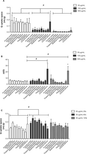
Figure 5 Capillary depletion results. The percentage of the ceria ENM dose in the capillary-rich fraction and brain-rich flow fraction for 100 μg/mL, at a rate of 20 mL/minute, 120-second perfusion duration, followed by 20-second washout.
Abbreviation: ENM, engineered nanomaterial.
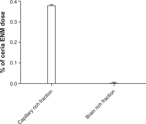
Figure 6 The capillaries in the (A) choroid plexus, (B) hippocampus (mid-CA1 region), and (C) pituitary were not affected by the high perfusate flow rate.
Note: Rats were perfused with 100 μg ceria ENM/mL, at a flow rate of 20 mL/minute, and perfusion duration of 120 seconds.
Abbreviation: ENM, engineered nanomaterial.
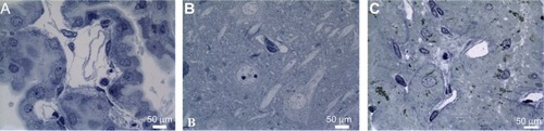
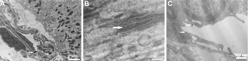
Figure 7 (A) A capillary with intact endothelial lining in the choroid plexus containing two ceria ENM agglomerates (arrows). (B) A vessel in the hippocampus and (C) a vessel from the pituitary gland associated with fine ceria ENM (arrows).
Note: Rats were perfused with 100 μg ceria ENM/mL, at a flow rate of 20 mL/minute, and perfusion duration of 120 seconds.
Abbreviation: ENM, engineered nanomaterial.