Figures & data
Figure 1 Gel electrophoresis of PEG-g-PEI-SPION/siRNA and scAbGD2-PEG-g-PEI-SPION/siRNA at various N/P ratios from 1.8 to 2.4.
Notes: The band intensities of nontargeting polyplexes. The brightness of EB-stained siRNA bands decreased with increasing the N/P ratio of nontargeting and targeting polyplexes. siRNA condensation by both PEG-g-PEI-SPION and scAbGD2-PEG-g-PEI-SPION completely retarded the siRNA motion by almost the same amount at an N/P ratio of 2.2.
Abbreviations: PEG-g-PEI-SPION, polyethylene glycol-grafted polyethylenimine superparamagnetic iron oxide nanoparticle; siRNA, small interfering ribonucleic acid; N/P, nitrogen-phosphorus; EB, ethidium bromide.
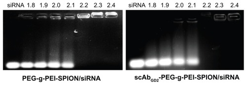
Figure 2 The cytotoxicities of PEG-g-PEI-SPION/siRNA and scAbGD2-PEG-g-PEI-SPION/ siRNA polyplexes in SK-N-SH cells.
Notes: The cytotoxicities of PEG-g-PEI-SPION/siRNA and scAbGD2-PEG-g-PEI-SPION/ siRNA polyplexes in SK-N-SH cells were evaluated using the CCK-8 assay. The siRNA concentration was 0.15 μg per well (in 300 μL cell culture medium), and the polyplex added into each well was adjusted according to the N/P ratios. There were no statistical differences between the two groups (P > 0.05). The results were expressed as the means ± SD (n = 3).
Abbreviations: PEG-g-PEI-SPION, polyethylene glycol-grafted polyethylenimine superparamagnetic iron oxide nanoparticle; siRNA, small interfering ribonucleic acid; CCK-8, cell counting kit-8; N/P, nitrogen-phosphorus; P, probability level; SD, standard deviation.
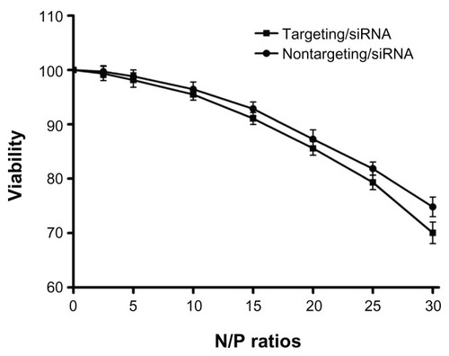
Figure 3 The particle size and zeta potential of PEG-g-PEI-SPION (nontargeting), scAbGD2-PEG-g-PEI-SPION (targeting), PEG-g-PEI-SPION/siRNA and scAbGD2-PEG-g-PEI-SPION/ siRNA polyplexes.
Abbreviations: PEG-g-PEI-SPION, polyethylene glycol-grafted polyethylenimine superparamagnetic iron oxide nanoparticle; siRNA, small interfering ribonucleic acid; N/P, nitrogen-phosphorus.
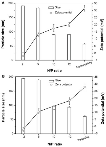
Figure 4 The transfection efficiency in SK-N-SH cells.
Notes: The transfection efficiency in SK-N-SH cells at various N/P ratios of PEG-g-PEI-SPION/siRNA (nontargeting), scAbGD2-PEG-g-PEI-SPION/siRNA (targeting) and scAbGD2-PEG-g-PEI-SPION/siRNA with free GD2 antibody (targeting/GD2). There were significant differences between targeting and nontargeting polyplexes.
Abbreviations: PEG-g-PEI-SPION, polyethylene glycol-grafted polyethylenimine superparamagnetic iron oxide nanoparticle; siRNA, small interfering ribonucleic acid; N/P, nitrogen-phosphorus.
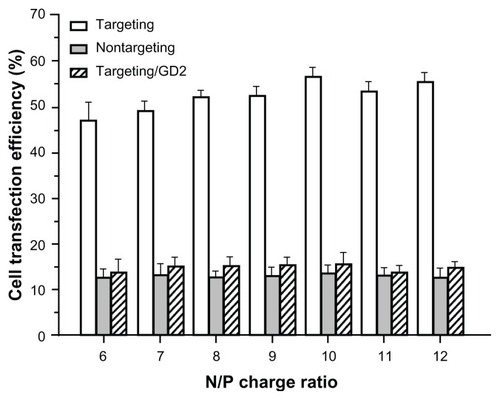
Figure 5 High magnification (100×) laser confocal microscopic images of SK-N-SH cells incubated for 2 hours with scAbGD2-PEG-g-PEI-SPION/siRNA (targeting) and PEG-g-PEI-SPION/siRNA (nontargeting).
Notes: SK-N-SH cells were incubated for 30 minutes with free GD2 antibody and then incubated with scAbGD2-PEG-g-PEI-SPION/siRNA (targeting/GD2) for 2 hours in cell culture medium. All polyplexes were formed at N/P = 10.
Abbreviations: PEG-g-PEI-SPION, polyethylene glycol-grafted polyethylenimine superparamagnetic iron oxide nanoparticle; siRNA, small interfering ribonucleic acid; N/P, nitrogen-phosphorus.

Figure 6 T2-weighted imaging of SK-N-SH cells. (A) T2-weighted imaging of SK-N-SH cells after 2-hour incubations with scAbGD2-PEG-g-PEI-SPION, PEG-g-PEI-SPION, and scAbGD2-PEG-g-PEI-SPION/GD2 at Fe concentrations of 5, 10, 20 and 40 μg/mL in DMEM. Untreated cells were used as the control group. Cells were scanned with a 1.5T MRI scanner. (B) shows the normalized MR intensity of different polymers at various Fe concentrations (n = 3).
Abbreviations: PEG-g-PEI-SPION, polyethylene glycol-grafted polyethylenimine superparamagnetic iron oxide nanoparticle; DMEM, Dulbecco’s modified eagle medium; MRI, magnetic resonance imaging; MR, magnetic resonance. with either nontargeting or targeting/GD2 polyplexes. The results are consistent with results obtained through RT-PCR and indicate that targeting polyplexes have a stronger suppressive effect on the Bcl-2 gene.
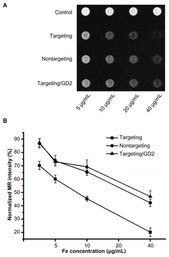
Figure 7 Cell apoptosis assessed by TUNEL at N/P = 10, the applied siRNA amount per well in cell culture was set constantly to 100 nM. (A) Images show cells incubated with: scAbGD2-PEG-g-PEI-SPION/siRNA (targeting), PEG-g-PEI-SPION/siRNA (nontargeting), scAbGD2-PEG-g-PEI-SPION/siRNA with free GD2 antibody (targeting/GD2). (B) Quantitative analysis of the percentage of apoptotic cells using a standard cell counting method in TUNEL assay (n = 3). When compared to scAbGD2-PEG-g-PEI-SPION/ siRNA (targeting), *P < 0.01 for both nontargeting and targeting/GD2 polyplexes.
Abbreviations: siRNA, small interfering ribonucleic acid; PEG-g-PEI-SPION, polyethylene glycol-grafted polyethylenimine superparamagnetic iron oxide nanoparticle; P, probability level.
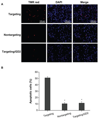
Figure 8 Efficiencies of PEG-g-PEI-SPION/siRNA, scAbGD2-PEG-g-PEI-SPION/ siRNA, and scAbGD2-PEG-g-PEI-SPION/siRNA with free GD2 antibody in suppressing Bcl-2 gene expression in SK-N-SH cells (N/P = 10). (A) Suppression of Bcl-2 mRNA levels as quantified by real-time RT-PCR (n = 3), When compared to scAbGD2-PEG-g-PEI-SPION/siRNA (targeting), *P < 0.01 for control, nontargeting and targeting/GD2 polyplexes. (B) Suppression of the protein expression of the Bcl-2 gene was evaluated by Western blot analysis.
Abbreviations: PEG-g-PEI-SPION, polyethylene glycol-grafted polyethylenimine superparamagnetic iron oxide nanoparticle; siRNA, small interfering ribonucleic acid; RT-PCR, reverse transcription polymerase chain reaction; P, probability level.
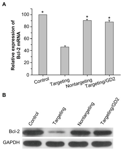
Figure 9 The average tumor size in different groups.
Notes: They include the PBS (control), PEG-g-PEI-SPION/siRNA (nontargeting), scAbGD2-PEG-g-PEI-SPION/siRNA (targeting). When compared to scAbGD2-PEG-g-PEI-SPION/siRNA (targeting). *P < 0.01 for both control and nontargeting polyplexes. All polyplexes were formed at N/P = 10.
Abbreviations: PBS, phosphate buffered saline; PEG-g-PEI-SPION, polyethylene glycol-grafted polyethylenimine superparamagnetic iron oxide nanoparticle; siRNA, small interfering ribonucleic acid; P, probability level; N/P, nitrogen-phosphorus.
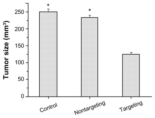
Figure 10 Immunohistological characteristics of SK-N-SH tumors (sections were immunostained with Bcl-2 monoclonal antibodies). The brown stains indicated Bcl-2 protein. (A and B) PBS control, (C and D) nontargeting polyplex therapy, (E and F) targeting polyplex therapy. The black arrow mark apoptotic cell with condensation or nuclei fragmentation.
Abbreviation: PBS, phosphate buffered saline.
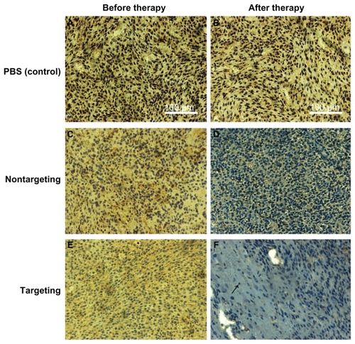
Figure 11 Tumor targeting evaluation in vivo. (A) Group 1: mice with SK-N-SH tumors after injection of PEG-g-PEI-SPION; Group 2: mice with SK-N-SH tumors after injection of scAbGD2-PEG-g-PEI-SPION; Group 3: mice with HepG2 tumors after injection of PEG-g-PEI-SPION; Group 4: mice with HepG2 tumors after injection of scAbGD2-PEG-g-PEI-SPION. (B) The normalized MR intensity of all groups after injection.
Abbreviations: MR, magnetic resonance; PEG-g-PEI-SPION, polyethylene glycol-grafted polyethylenimine superparamagnetic iron oxide nanoparticle.
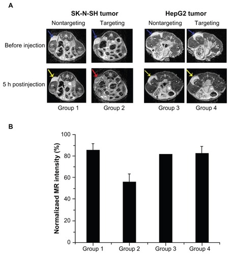
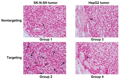
Figure 12 Prussian blue staining was performed on histological sections of tumors.
Note: Blue positive particles marked by black arrows.