Figures & data
Figure 1 Schematic of composite scaffold preparation and groups.
Abbreviations: β-TCP, β-tricalcium phosphate; DOX, doxorubicin hydrochloride; PCL, polycaprolactone.
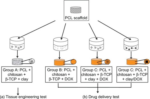
Table 1 Scaffold composition in different groups
Figure 2 Cumulative DOX release (%) from Group B (DOX without clay scaffold), Group C (DOX with clay scaffold), and Group D (Clay/DOX carrier scaffold) at 37°C in PBS (pH 7.4) for 2 months.
Abbreviations: PCL, polycaprolactone; β-TCP, β-tricalcium phosphate; DOX, doxorubicin hydrochloride; PBS, phosphate-buffered saline.
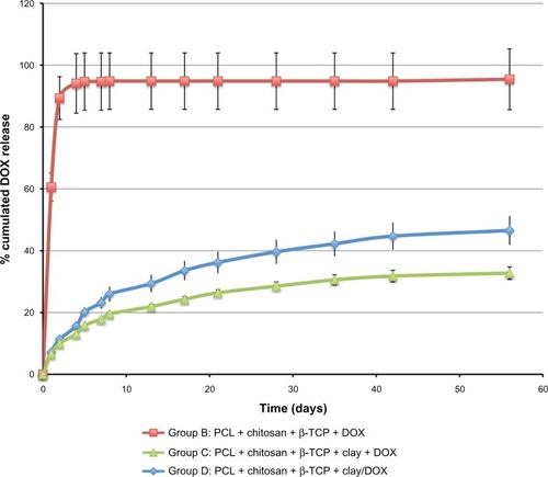
Figure 3 Cell adhesion, viability, and proliferation in the scaffolds. (A) SEM images of cellular scaffolds from the cell seeding side on Days 1, 7, 14, and 21. The arrows point to the cells on Day 1 and to the crystal-like extracellular matrix deposition on Day 21. EDX analysis of the element components revealed the mineralized nodules as calcium phosphate salts, consisting of P, Ca, and O elements in the cell culture scaffold for Day 21, compared to day 0 in the cell free scaffold. (B) Confocal micrographs of cell viability on Days 1, 7, 14, and 21. Green pixels: live cells; red pixels: nuclei; green and red pixel overlap: chitosan structure. Upper panels show projections of a 150 μm optical section onto a single plane (magnification 5×). Bottom panels show single planes at magnification 20×. (C) Quantification of dsDNA in cellular scaffolds by means of PicoGreen assay on Days 1, 7, 14, and 21.
Notes: The amount of DNA is expressed as mean ± SD (n = 4). Different letters (a–c) indicate significant differences (P < 0.05) between sampling days.
Abbreviations: ALP, alkaline phosphatase; SEM, scanning electron microscopy; EDX, energy dispersive X-ray spectrometer.
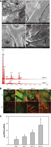
Figure 4 Osteogenic differentiation and mineralization of cellular scaffolds. (A) Activity of the ALP enzyme on Days 1, 7, 14, and 21. The activity is expressed as mean ± SD (n = 4). Activity is indicated in nanomole p-nitrophenol/microgram DNA per minute (nmol/μg DNA * min). Different letters (a–c) indicate significant differences (P < 0.05) between sampling days. (B) ALP staining. (C) Calcium contents per scaffold on Days 1, 7, 14, and 21. The amount of calcium is expressed as mean ± SD (n = 4). Different letters (a–c) indicate significant differences (P < 0.05) between sampling days. (D) von Kossa staining of scaffolds.
Abbreviations: ALP, alkaline phosphatase; SD, standard deviation.
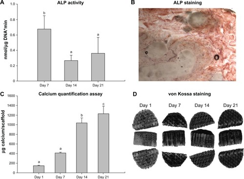
Figure 5 Histology results. (A) Representative histology micrographs of H&E stain showing cross-sections of the top part of cell/scaffold constructs after 7-day and 21-day culture. Bars = 1 mm. Nuclei are stained dark blue (basophilic), ECM and cytoplasm are stained purple, and the chitosan foam is stained orange. (B) Immunohistology staining of anti-human osteocalcin of the scaffolds after 21-day culture. Sections stained without anti-human osteocalcin served as control.
Note: Arrows point to representative positive staining of osteocalcin.
Abbreviations: OC, osteocalcin; H&E, hematoxylin and eosin; ECM, extracellular matrix.
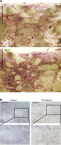
Figure 6 Principle of the composite scaffold with drug eluting matrix embedding. (A) Following tumor resection, remnant tumor cells (orange) may be present and there is an increased risk of recurrence. (B) Implantation of mechanically stable porous scaffold with controlled drug release functions will provide immediate load bearing support and localized anti-cancer treatment. (C) After all the drug has been released, the implant will aid in tissue regeneration of the wound site.
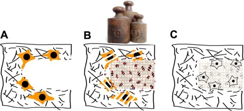
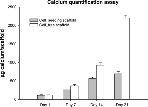
Figure 7 Calcium deposition contents per scaffold on cell-free scaffolds in comparison with cell-seeded scaffolds on Days 1, 7, 14, and 21.