Figures & data
Figure 1 Schematic diagram of the chemical structure and the reaction of Cys-penetratin and MPB-PE used to prepare penetratin-conjugated liposomes.
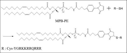
Table 1 Physical Characteristics of the Liposome Formulations
Figure 2 TEM images of the control liposomes (A) and the penetratin-conjugated liposomes (B).
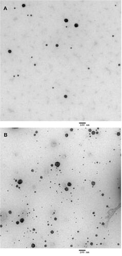
Figure 3 Cytotoxicity of various liposome concentrations in TR146 cells after 24 hours of incubation. Error bars represent SD (n = 5). ***p < 0.001 versus control (no treated group).
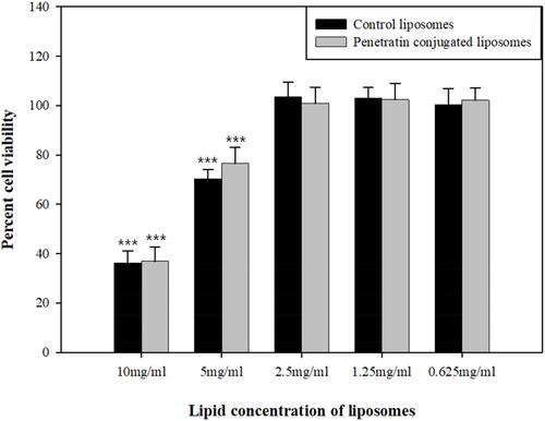
Figure 4 Fluorescence intensities of Alexa 647-sCT after two hours of treatment with the control and penetratin-conjugated liposomes as determined using flow cytometry. Representative fluorescence intensity (A) and relative MFI values of Alexa 647-sCT (B). All data represent the mean ± SD (n = 3). ***p<0.001 vs sCT, ###p<0.001 vs Control liposomes.
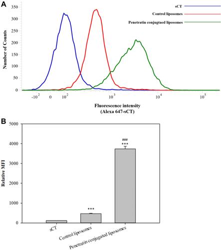
Figure 5 Confocal laser scanning microscopy (CLSM) images of the control liposomes containing Alexa 647-sCT and penetratin-conjugated liposomes in TR146 cells.
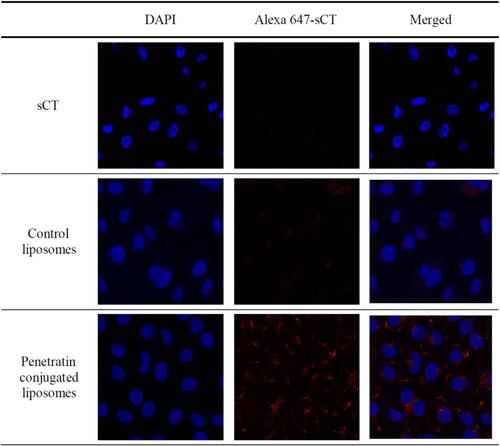
Table 2 TEER Values of the Formulations Before and After Permeability Experiments Using TR 146 Cell Layers
Figure 6 In vitro TR146 cell permeation profiles of sCT, control liposomes, and penetratin-conjugated liposomes.
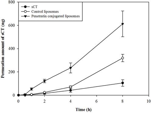
Table 3 Permeation Parameters Calculated from the TR146 Cell Permeation Study
Table 4 Permeation Parameters Calculated from the Buccal Tissue Permeation Study
Figure 7 Ex vivo buccal tissue permeation profiles of sCT, control liposomes, and penetratin-conjugated liposomes.
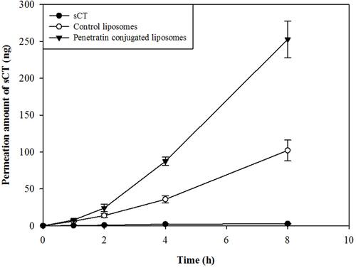
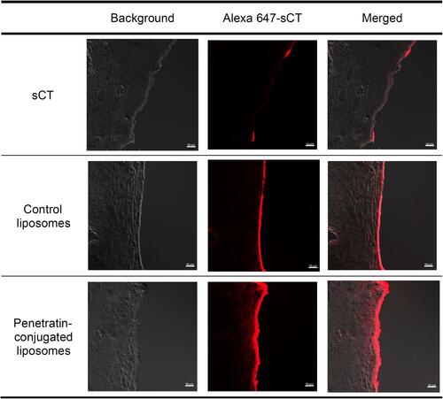
Figure 8 Confocal laser scanning microscopy (CLSM) images of the control liposomes containing Alexa 647-sCT and the penetratin-conjugated liposomes after permeation through the buccal tissue at 8 h.