Figures & data
Figure 1 Synthetic scheme of LTVSPWY-PEG-CS.
Abbreviations: PEG, poly(ethylene glycol); CS, chitosan.
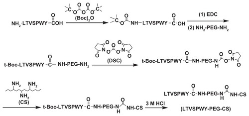
Figure 2 1H NMR spectra of LTVSPWY-PEG-CS (A), peptide LTVSPWY (B), CS (C) and NH2-PEG-NH2 (D). The important peaks are pointed out.
Abbreviations: PEG, poly(ethylene glycol); CS, chitosan.
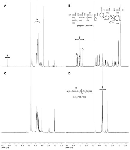
Table 1 Size and zeta potential of magnetic nanoparticles
Figure 3 Characteristics of magnetic nanoparticles. Size distribution (A) and zeta potential (B) of LTVSPWY-PEG-CS-modified magnetic nanoparticles obtained by dynamic light scattering.
Abbreviations: PEG, poly(ethylene glycol); CS, chitosan.
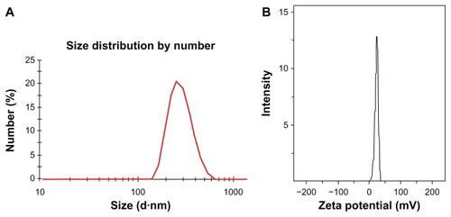
Figure 4 Transmission electron microscopic images of magnetic nanoparticles. (A) LTVSPWY-PEG-CS-modified magnetic nanoparticles, (B) PEG-CS-modified magnetic nanoparticles, and (C) CS-modified magnetic nanoparticles.
Note: The bar is 50 nm.
Abbreviations: PEG, poly(ethylene glycol); CS, chitosan.
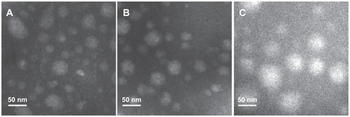
Figure 5 Cytotoxicity of magnetic nanoparticles against A549 (A) and SKOV-3 (B) cells. The magnetic nanoparticles prepared consisted of 50 μg/mL Fe3O4, 50 μg/mL oleic acid, 350 μg/mL monostearin, and 50 μg/mL chitosan.
Note: Data represent the mean ± standard deviation (n = 3).
Abbreviations: PEG, poly(ethylene glycol); CS, chitosan.
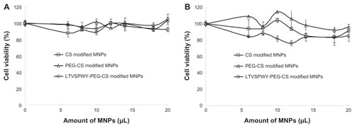
Figure 6 Fluorescence images of competitive cellular uptake of RITC-labeled magnetic nanoparticles for 12 hours. A549 cells (HER2-negative, cytoplasmic membrane-labeled with PKH67 fluorescent linker, green) cocultured with HER2-overexpressing SKOV-3 cells were incubated with RITC-labeled magnetic nanoparticles (red). (A) LTVSPWY-PEG-CS-modified magnetic nanoparticles, (B) PEG-CS-modified magnetic nanoparticles, and (C) chitosan-modified magnetic nanoparticles.
Note: The cells were all stained with Hoechst 33342.
Abbreviations: RITC, rhodamine B isothiocyanate; PEG, poly(ethylene glycol); CS, chitosan.
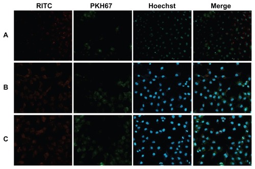
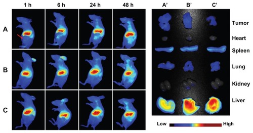
Figure 7 In vivo and ex vivo biodistribution of DiR-loaded magnetic nanoparticles after intravenous tail injection in tumor-bearing nude mice. The fluorescent images of in vivo tumor-bearing nude mice (A–B) and ex vivo image of major organs (A′–C′) using DiR probes with three different magnetic nanoparticles, ie, (A and A′) LTVSPWY-PEG-CS-modified magnetic nanoparticles, (B and B′) PEG-CS-modified magnetic nanoparticles, and (C and C′) chitosan-modified magnetic nanoparticles.
Notes: The images were spectrally resolved. The tumor area is emphasized by a red arrow.
Abbreviations: PEG, poly(ethylene glycol); CS, chitosan. for the development of novel molecular imaging probes in the early detection and diagnosis of cancer.