Figures & data
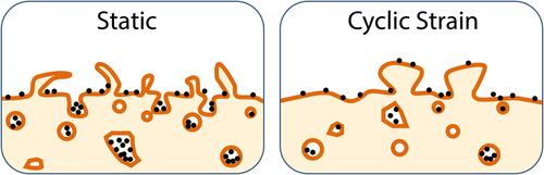
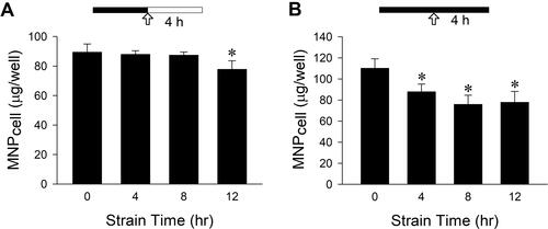
Figure 1 VSMC uptake of PAA-MNPs in response to mechanical strain with different strategies. Cyclic strain was applied as indicated by the solid bar for 4–12 h before or during incubation of (A–C) 200-nm or (D) 50-nm PAA-MNPs, or PLL-MNPs with (E) positive and (F) negative ζ-potentials at 100 μg/mL (25 μg/cm2) with VSMCs. Cell-associated MNPs (MNPcell or percentage of control) was measured 4 h after administration of MNPs (A, C-F) or 4 hr after application of cyclic strain (B). The arrows indicate the MNP administration; filled vs unfilled bars represent incubation with or without cyclic strain, respectively. Values are means ± SEM (n=3). *p<0.05 compared with corresponding values without strain.
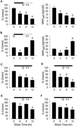
Figure 2 Changes in plasma membrane morphology were associated with reduced particle internalization in response to cyclic strain. VSMCs were subjected to cyclic strain for total of 12 h (A and B) with PAA-MNPs (100 µg/mL; 25 µg/cm2) added 8 h after initiation of strain (B and C). Cyclic strain-induced morphology changes from filopodia (B; left) to blunted protrusion (B; right) in the presence of PAA-MNPs are representative of 15 and 14 cells, respectively. The bottom panels (B) are amplifications of the areas in the rectangles. Quantitative analysis of intracellular (C) vesicle numbers, (D) vesicle size, and (E) areas of cells containing iron in TEM images of cells subjected to strain (n = 20) or without strain (control; n = 23). Values are means ± SEM. *p <0.05 compared with the corresponding control.
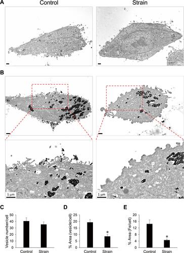
Figure 3 Representative SEM images of VSMCs subjected to mechanical strain during incubation with PAA-MNPs. (A) VSMCs were subjected to cyclic strain for 12 h or in steady culture conditions (control). (B) Cyclic strain was applied for 8 h before administration of PAA-MNPs (25 μg/mL; 6.25 μg/cm2), followed by additional 4 h of strain. The right panels are amplification of the areas as indicated on the left in (A and B). Scale bars indicate 1 μm. (C) SEM/EDS surface elemental analysis was conducted in the indicated areas (ROI 1 & 2) of the surface of VSMCs exposed to PAA-MNPs in static conditions (Bold values).
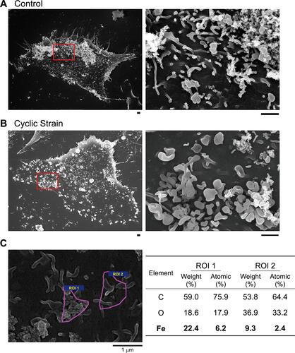
Figure 4 Representative confocal images of PAA-MNPs uptake by VSMCs subjected to cyclic strain. (A) VSMCs were incubated with PAA-MNPs (100 µg/mL; 25 µg/cm2, green) in culture dishes coated with fibronectin for 4 h; (B) VSMCs were subjected to cyclic strain for 8 h before administration of PAA-MNPs, followed by additional 4 h of cyclic strain with PAA-MNPs in the culture media. Cell membrane, nuclei and focal adhesions were stained by WGA (red), DAPI (blue), and mouse anti-vinculin antibody (white), respectively.
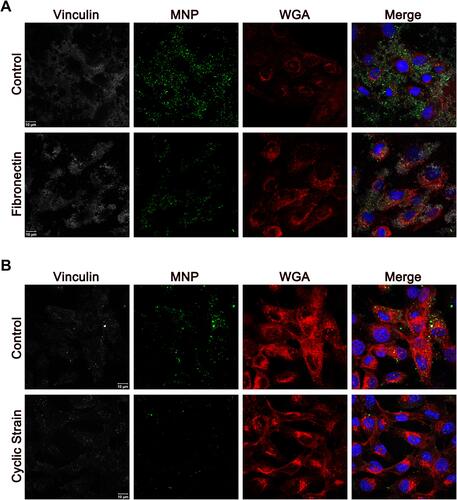
Figure 5 Cytochalasin D, but not blebbistatin, reduced VSMC uptake of PAA-MNPs in a concentration-dependent manner. Confocal images of VSMCs with 1-h pretreatment of (A) cytochalasin D or (B) blebbistatin at different concentrations. The cytoskeleton and nuclei were stained by phalloidin (red) and DAPI (blue), respectively. VSMCs were pretreated with cytochalasin D (C) or blebbistatin (D) for 1 h followed by incubation with PAA-MNPs (100 µg/mL; 25 µg/cm2) for 3 h prior to measurement. Values are means ± SEM (n=4). *p <0.05 compared with the control value.
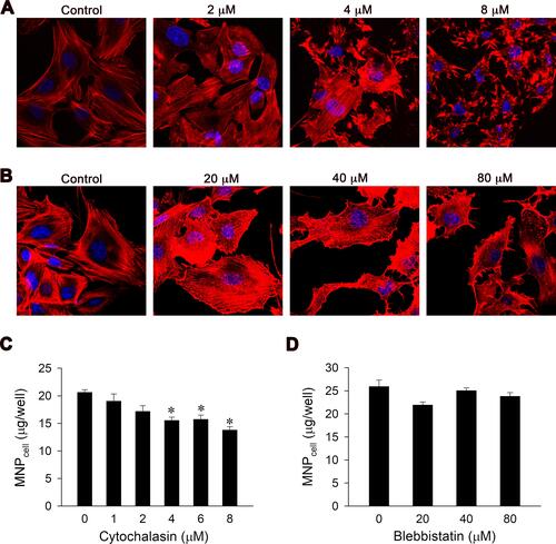
Figure 6 Mechanical strain attenuated PAA-MNPs uptake by LN-229 cells with time. Mechanical strain was applied as indicated by the solid bar for 4–12 h (A) before or (B) during incubation of LN-229 cells with PAA-MNPs (100 µg/mL; 25 µg/cm2). The arrows indicate the MNP administration; filled vs unfilled bars represent incubation with or without cyclic strain, respectively. Values are means ± SEM (n = 3). *p <0.05 compared with corresponding values without strain.