Figures & data
Figure 1 Single- and dual-color targeted SPNs. (A) Schematic representation of SPNs characterized by a COOH-derivatized PF-127 shell embedded in a silica/dye core. For each sample of single- and dual-color SPNs, the corresponding absorption and emission spectra in water solution are reported. (B) CGIYRLRS and CGVYSLRS peptides were selected by phage display biopanning of random libraries on matched hepatic metastasis/normal liver tissue pairs from patients with advanced CRC, and were demonstrated to be specific ligands for the metastatic tissue.
Abbreviations: CRC, colorectal cancer; PF-127, Pluronic®F127; SPN, silica-poly(ethylene glycol) nanoparticles.
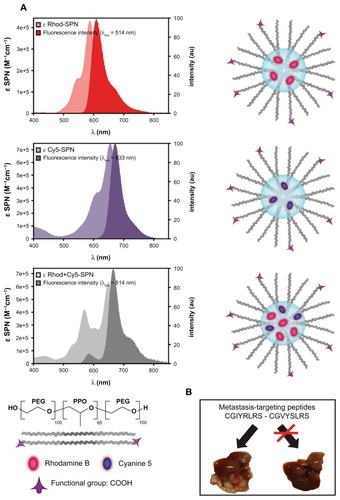
Figure 2 Targeted SPNs recognize human hepatic metastases ex vivo. Frozen 10-μm sections of (A) matched (grossly) normal liver, (B) hepatic metastasis, (C) (grossly) normal colon, and (D) primary tumor were fixed in 4% formaldehyde, before incubation with either untargeted, CGIYRLRS-, or CGVYSLRS-(Rhod+Cy5)-SPNs for 4 h at RT. After washing, the SPN-emitted fluorescence was analyzed using confocal microscopy keeping all parameters constant, and the output was converted into a false color LUT Fire for clear visualization. Colocalized pixels were identified using ImageJ software. Experiments were performed, giving similar results, on specimens from ten patients with metastatic CRC; exemplary images from tissues of patient #P85 are shown. (E) Quantification of SPN binding is expressed as the intensity of emitted pixel following excitation at 514 nm (Rhod) and 633 nm (Cy5), and calculated as a mean value of five images for each tissue. Cell nuclei were stained with 4′,6-diamidino-2-phenylindole and are visualized in blue. All tissue slices were imaged in the same way.
Abbreviations: CRC, colorectal cancer; Rhod, rhodamine; RT, room temperature; SPN, silica-poly(ethylene glycol) nanoparticles.
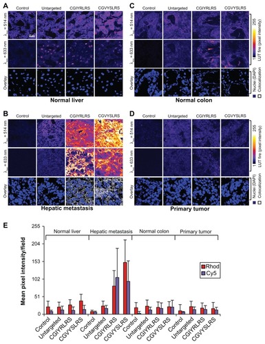
Figure 3 Targeted SPNs home to hepatic metastases in vivo. NOD/SCID mice bearing a primary spleen tumor and multiple liver metastases were injected intravenously with single- [control (A, Rhod; B, Cy5) or CGIYRLRS- (D, Rhod; E, Cy5)] or dual-color [control (C) or CGIYRLRS (F)] SPNs. After 16 h, mice were euthanized and the explanted organs were photographed in toto using high-resolution CCD cameras connected to a fluorescence stereomicroscope. In d, e, and f, orange arrows indicate blood vessels crossing the hepatic metastasis; in e and f, white arrows indicate submillimetric metastatic foci. Samples of the same tissues were OCT-frozen, cut into 10-μm slices, and evaluated at a micrometric scale by confocal analysis of the single- [control (G, Rhod; I, Cy5), CGIYRLRS (H, Rhod; J, Cy5)] or dual-color [control (K) or CGIYRLRS (L)] SPN-emitted fluorescence. To visualize blood vessels, staining for CD31 was superimposed on the SPNs signal, and was visualized using secondary antibodies Alexa Fluor®647 (G and H) and Alexa Fluor®488 (I and J), for overlay with (Rhod)-SPNs and (Cy5)-SPNs, respectively. In the case of dual-color nanoparticles, a further control is visualized, ie, samples of primary tumors from mice injected with either control (M) or CGIYRLRS (N) SPNs. Microphotographs G–J and M–N refer to a single tissue type (metastasis and primary tumor, respectively); microphotographs k and l depict the interface between normal liver and hepatic metastasis; in this second case, a dotted line has been added to separate the two tissues, as indicated on the photo. Fifty to 80 series of images superimposing CD31 were stained with either (Cy5)-SPNs (O, particular of the field in J) or (Rhod+Cy5)-SPNs (P, particular of the field in L, in which CD31 was visualized using the secondary antibody DyLightTM405). SPNs were composed to reconstruct 3D models using IMARIS.
Abbreviations: NOD/SCID, nonobese diabetic/severe combined immunodeficient; OCT, optimal cutting temperature; SPN, silica-poly(ethylene glycol) nanoparticles.
![Figure 3 Targeted SPNs home to hepatic metastases in vivo. NOD/SCID mice bearing a primary spleen tumor and multiple liver metastases were injected intravenously with single- [control (A, Rhod; B, Cy5) or CGIYRLRS- (D, Rhod; E, Cy5)] or dual-color [control (C) or CGIYRLRS (F)] SPNs. After 16 h, mice were euthanized and the explanted organs were photographed in toto using high-resolution CCD cameras connected to a fluorescence stereomicroscope. In d, e, and f, orange arrows indicate blood vessels crossing the hepatic metastasis; in e and f, white arrows indicate submillimetric metastatic foci. Samples of the same tissues were OCT-frozen, cut into 10-μm slices, and evaluated at a micrometric scale by confocal analysis of the single- [control (G, Rhod; I, Cy5), CGIYRLRS (H, Rhod; J, Cy5)] or dual-color [control (K) or CGIYRLRS (L)] SPN-emitted fluorescence. To visualize blood vessels, staining for CD31 was superimposed on the SPNs signal, and was visualized using secondary antibodies Alexa Fluor®647 (G and H) and Alexa Fluor®488 (I and J), for overlay with (Rhod)-SPNs and (Cy5)-SPNs, respectively. In the case of dual-color nanoparticles, a further control is visualized, ie, samples of primary tumors from mice injected with either control (M) or CGIYRLRS (N) SPNs. Microphotographs G–J and M–N refer to a single tissue type (metastasis and primary tumor, respectively); microphotographs k and l depict the interface between normal liver and hepatic metastasis; in this second case, a dotted line has been added to separate the two tissues, as indicated on the photo. Fifty to 80 series of images superimposing CD31 were stained with either (Cy5)-SPNs (O, particular of the field in J) or (Rhod+Cy5)-SPNs (P, particular of the field in L, in which CD31 was visualized using the secondary antibody DyLightTM405). SPNs were composed to reconstruct 3D models using IMARIS.Abbreviations: NOD/SCID, nonobese diabetic/severe combined immunodeficient; OCT, optimal cutting temperature; SPN, silica-poly(ethylene glycol) nanoparticles.](/cms/asset/c752793a-9492-451a-a1e1-e6ec3bab93b7/dijn_a_33825_f0003_c.jpg)
Figure S1 Characterization of the fluorescent SPNs. (A) TEM analysis of SPN silica cores for the evaluation of size distribution. (B) Dynamic light scattering distribution of the nanoparticle hydrodynamic diameter. Exemplary graphs referred to CGIYRLRS-(Rhod)-SPNs are shown.
Abbreviations: SPN, silica-poly(ethylene glycol) nanoparticles; TEM, transmission electron microscopy.
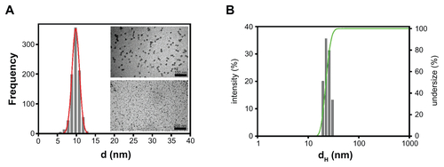
Figure S2 Single-color targeted SPNs recognize human hepatic metastases ex vivo Frozen sections (10-μm) of matched (grossly) normal liver and hepatic metastasis were fixed in 4% formaldehyde before being incubated with 5 × 10¹² of either untargeted, CGIYRLRS-, or (A) CGVYSLRS-(Rhod)- or (B) (Cy5)-SPNs for 4 h at RT. After 3 washes in TBS-T, the SPN-emitted fluorescence was analyzed by confocal microscopy, and the output was converted in a false color LUT Fire scale for prompt visualization. The experiment was performed on specimens from 10 patients with metastatic CRC; exemplary images from tissues of patient #P85 are shown.
Abbreviations: CRC, colorectal cancer; RT, room temperature; SPN, silica-poly(ethylene glycol) nanoparticles; TBS-T, Tris-buffered saline containing Tween-20.
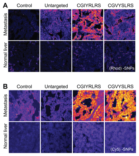
Table S1 Morphological and photophysical properties of the SNPs and of their doping dyes