Figures & data
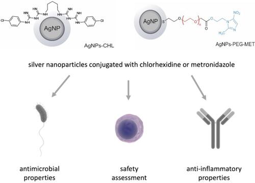
Figure 1 UV-VIS spectra of AgNPs-CHL and AgNPs-PEG-MET recorded in water solutions.
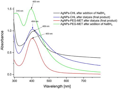
Figure 2 TEM micrographs of the synthesized NPs: (A) AgNPs-CHL; (B) AgNPs-PEG-MET; (C) AgNPs-CHL; (D) AgNPs-PEG-MET. The histograms were prepared based on these images.
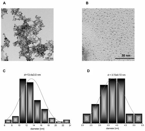
Figure 3 The number averaged hydrodynamic diameter distribution of the synthesized NPs obtained from DLS measurements.
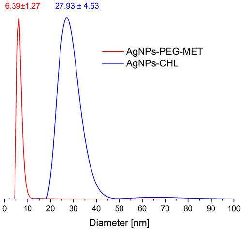
Figure 4 TGA curves recorded during the heating AgNPs-CHL and CHL not attached on the silver surface under nitrogen atmosphere (solid lines) and their derivatives with temperature (dotted lines with the same colours).
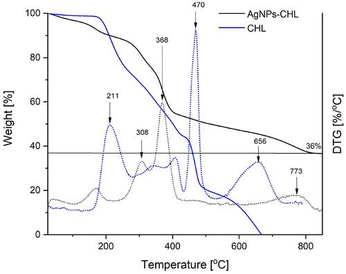
Figure 5 TGA curves recorded during the heating of AgNPs-PEG-MET. MET is metronidazole not attached to the silver surface, and PEG is a derivative used to link the silver and the drug. The measurements were taken under a nitrogen atmosphere (solid lines); derivatives of the curves with temperature are represented as dotted lines with the same colours.
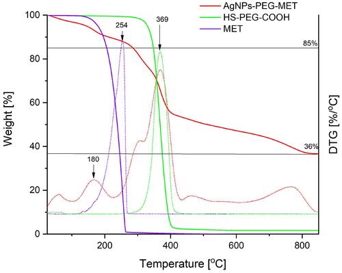
Table 1 Minimum Inhibitory Concentrations (µg/mL) Against Tested Strains
Table 2 Minimum Biofilm Eradication Concentrations (µg/mL) Against Tested Strains
Figure 6 Ultrastructure of S. maltophilia and S. mutans after treatment with AgNPs for 24 hours: (A) S. maltophilia control; (B) S. maltophilia treated with AgNPs-CHL; (C) S. maltophilia treated with AgNPs-PEG-MET; (D) S. mutans control; (E) S. mutans treated with AgNPs-CHL; (F) S. mutans treated with AgNPs-PEG-MET. The scale bar is present on the left side of each picture. Arrows indicate NPs.
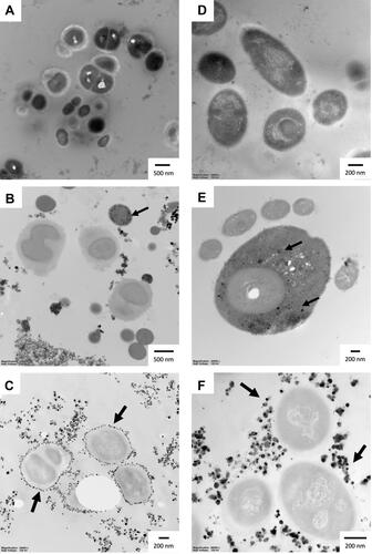
Table 3 Half Maximal Inhibitory Concentration (IC50) for AgNPs-CHL and AgNPs-PEG-MET
Table 4 Mean SI – Selectivity index (Mean IC50/MIC) for AgNPs-CHL and AgNPs-PEG-MET. The Mean IC50 Value for HGF-1 Cells () and MIC Values () Were Used in These Calculations. Values Were Rounded to the Nearest Decimal
Figure 7 Ultrastructure of the hFOB1.19 cells after 24 hours of incubation with AgNPs: (A and B) control cells; (C and D) cells treated with 1 µg/mL AgNPs-CHL; (E and F) cells treated with 1 µg/mL AgNPs-PEG-MET. Arrows indicate NPs. *Autophagic vacuoles; V–vacuoles; N – nucleus; M – mitochondria. The scale bar is present on the left side of each picture.
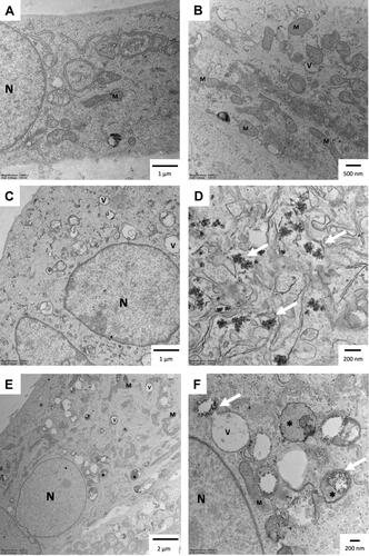
Figure 8 Effect of AgNPs-CHL and AgNPs-PEG-MET on cytokine secretion by RAW264.7 cells to medium. Cells were treated with AgNPs-drugs for 24 hours and LPS (1 µg/mL) was used to induce inflammation. Secretions: (A) IL-1β; (B) IL-6; (C) TNFα; (D) IL-8. Data are presented as mean ± SD. ###P < 0.001 (vs untreated control), ***P < 0.001 (vs control treated with LPS).
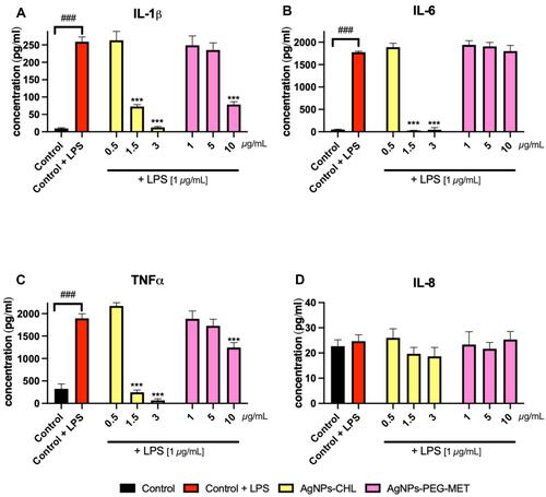
Figure 9 Effect of AgNPs-CHL and AgNPs-PEG-MET on intracellular production of cytokines by RAW264.7 cells. Cells were treated with AgNPs-drugs for 24 hours and LPS (1 µg/mL) was used to induce inflammation: (A) production of IL-1β; (B) production of IL-6; (C) production of TNFα; (D) production of IL-8. Data are presented as mean ± SD. ##P < 0.01 (vs untreated control), *P < 0.05, **P < 0.01, ***P < 0.001 (vs control treated with LPS).
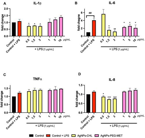
Figure 10 Effect of AgNPs-CHL and AgNPs-PEG-MET on intracellular production of reactive oxygen species in RAW264.7 cells. Cells were treated with AgNPs-drugs for 24 hours and LPS (1 µg/mL) was used to induce inflammation. Data are presented as mean ± SD. *P < 0.05, **P < 0.01 (vs unstimulated control).
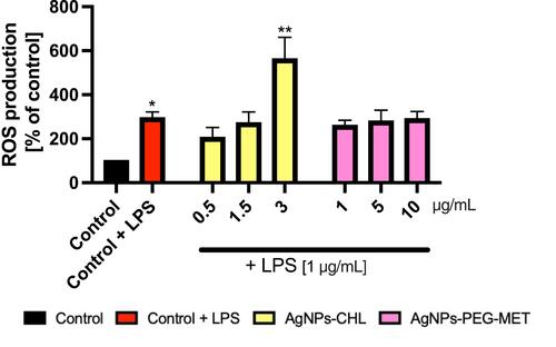
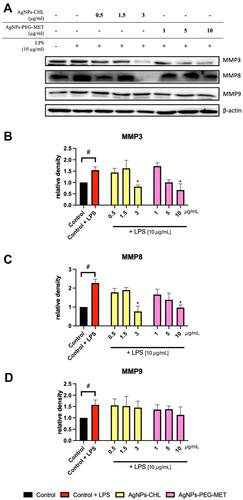
Figure 11 Effect of AgNPs-CHL and AgNPs-PEG-MET on metalloproteinase levels in hFOB1.19 cells. Cells were treated with AgNPs-drugs for 24 hours and LPS (10 µg/mL) was used to induce inflammation: (A) representative Western blots; (B) quantification of MMP3 levels; (C) quantification of MMP8 levels; (D) quantification of MMP9 levels. Data are presented as mean ± SD. #P<0.05 (vs unstimulated control), *P < 0.05 (vs control treated with LPS).