Figures & data
Figure 1 Schematic drawing of microsphere preparation.
Abbreviations: PLGA, poly(lactic-co-glycolic acid); MS, microspheres.
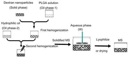
Figure 2 (A–D) Scanning electron micrographs of different samples. (A) Dextran nanoparticles; (B) dextran nanoparticles recovered from the prepared dextran nanoparticle–loaded microspheres using S/O/O/W; (C) control microspheres (prepared microspheres using W/O/W); (D) prepared dextran nanoparticle–loaded microspheres using S/O/O/W.
Abbreviations: S/O/O/W, solid-in-oil-in-oil-in-water; W/O/W, water-in-oil-in-water.
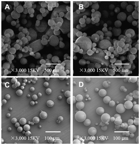
Figure 3 Monomer content of recovery G-CSF from different samples during preparation processes by SEC-HPLC (n = 5).
Notes: Samples: original G-CSF (G-CSF:dextran = 1.4:5.0 ± 0.3 mg) solution; G-CSF from dextran aqueous phase/polyethyene glycol aqueous phase emulation; G-CSF from dextran nanoparticles; G-CSF from prepared microspheres using the S/O/O/W method (PLGA 50/50 2A: 100.0 ± 0.5 mg PLGA, dextran nanoparticles [G-CSF:dextran, 1:4] = 10.0 ± 0.3 mg); G-CSF from control microspheres (prepared microspheres using the W/O/W method) (PLGA 50/50 2A: 100.0 ± 0.5 mg, G-CSF solution [G-CSF:dextran = 1:4] = 10.0 ± 0.3 mg). *P > 0.05; **P < 0.05.
Abbreviations: G-CSF, granulocyte colony–stimulating factor; SEC-HPLC, size-exclusion chromatography–high-pressure liquid chromatography; PLGA, polylactic-co-glycolic acid; S/O/O/W, solid-in-oil-in-oil-in-water; W/O/W, water-in-oil-in-water.
![Figure 3 Monomer content of recovery G-CSF from different samples during preparation processes by SEC-HPLC (n = 5).Notes: Samples: original G-CSF (G-CSF:dextran = 1.4:5.0 ± 0.3 mg) solution; G-CSF from dextran aqueous phase/polyethyene glycol aqueous phase emulation; G-CSF from dextran nanoparticles; G-CSF from prepared microspheres using the S/O/O/W method (PLGA 50/50 2A: 100.0 ± 0.5 mg PLGA, dextran nanoparticles [G-CSF:dextran, 1:4] = 10.0 ± 0.3 mg); G-CSF from control microspheres (prepared microspheres using the W/O/W method) (PLGA 50/50 2A: 100.0 ± 0.5 mg, G-CSF solution [G-CSF:dextran = 1:4] = 10.0 ± 0.3 mg). *P > 0.05; **P < 0.05.Abbreviations: G-CSF, granulocyte colony–stimulating factor; SEC-HPLC, size-exclusion chromatography–high-pressure liquid chromatography; PLGA, polylactic-co-glycolic acid; S/O/O/W, solid-in-oil-in-oil-in-water; W/O/W, water-in-oil-in-water.](/cms/asset/beff1a2c-5404-4def-bdf7-43263b81bbfe/dijn_a_33993_f0003_b.jpg)
Figure 4 Bioactivity from different samples during preparation processes (n = 5).
Notes: Samples: original G-CSF (G-CSF:dextran = 1.4:5.0 ± 0.3 mg) solution; G-CSF from dextran aqueous phase/polyethylene glycol aqueous phase emulation; G-CSF from dextran nanoparticles; G-CSF from prepared microspheres using the S/O/O/W method (PLGA 50/50 2A: 100.0 ± 0.5 mg PLGA, dextran nanoparticles [G-CSF:dextran, 1:4] = 10.0 ± 0.3 mg); G-CSF from control microspheres (prepared microspheres using the W/O/W method) (PLGA 50/50 2A: 100.0 ± 0.5 mg, G-CSF solution [G-CSF:dextran = 1:4] = 10.0 ± 0.3 mg). *P > 0.05; **P < 0.05.
Abbreviations: G-CSF, granulocyte colony–stimulating factor; PLGA, polylactic-co-glycolic acid; S/O/O/W, solid-in-oil-in-oil-in-water; W/O/W, water-in-oil-in-water.
![Figure 4 Bioactivity from different samples during preparation processes (n = 5).Notes: Samples: original G-CSF (G-CSF:dextran = 1.4:5.0 ± 0.3 mg) solution; G-CSF from dextran aqueous phase/polyethylene glycol aqueous phase emulation; G-CSF from dextran nanoparticles; G-CSF from prepared microspheres using the S/O/O/W method (PLGA 50/50 2A: 100.0 ± 0.5 mg PLGA, dextran nanoparticles [G-CSF:dextran, 1:4] = 10.0 ± 0.3 mg); G-CSF from control microspheres (prepared microspheres using the W/O/W method) (PLGA 50/50 2A: 100.0 ± 0.5 mg, G-CSF solution [G-CSF:dextran = 1:4] = 10.0 ± 0.3 mg). *P > 0.05; **P < 0.05.Abbreviations: G-CSF, granulocyte colony–stimulating factor; PLGA, polylactic-co-glycolic acid; S/O/O/W, solid-in-oil-in-oil-in-water; W/O/W, water-in-oil-in-water.](/cms/asset/35ba2594-46bf-49c0-850d-3731c4bdcd78/dijn_a_33993_f0004_b.jpg)
Figure 5 In vitro release profiles of microspheres (n = 5, P < 0.05).
Notes: ⋄: Prepared microspheres using the S/O/O/W method (PLGA 50/50 2A: 100.0 ± 0.5 mg PLGA, dextran nanoparticles (G-CSF:dextran, 1:4) = 10.0 ± 0.3 mg); ♦: control microspheres (prepared microspheres using W/O/W method) (PLGA 50/50 2A: 100.0 ± 0.5 mg, G-CSF solution [G-CSF:dextran = 1:4] = 10.0 ± 0.3 mg).
Abbreviations: G-CSF, granulocyte colony–stimulating factor; PLGA, polylactic-co-glycolic acid; S/O/O/W, solid-in-oil-in-oil-in-water; W/O/W, water-in-oil-in-water.
![Figure 5 In vitro release profiles of microspheres (n = 5, P < 0.05).Notes: ⋄: Prepared microspheres using the S/O/O/W method (PLGA 50/50 2A: 100.0 ± 0.5 mg PLGA, dextran nanoparticles (G-CSF:dextran, 1:4) = 10.0 ± 0.3 mg); ♦: control microspheres (prepared microspheres using W/O/W method) (PLGA 50/50 2A: 100.0 ± 0.5 mg, G-CSF solution [G-CSF:dextran = 1:4] = 10.0 ± 0.3 mg).Abbreviations: G-CSF, granulocyte colony–stimulating factor; PLGA, polylactic-co-glycolic acid; S/O/O/W, solid-in-oil-in-oil-in-water; W/O/W, water-in-oil-in-water.](/cms/asset/43f1ad78-06bf-4be1-b71d-04d847733dda/dijn_a_33993_f0005_b.jpg)
Figure 6 In vitro release relative bioactivity of microspheres (n = 5, P < 0.05).
Notes: ⋄: Prepared microspheres using the S/O/O/W method (PLGA 50/50 2A: 100.0 ± 0.5 mg PLGA, dextran nanoparticles [G-CSF:dextran, 1:4] = 10.0 ± 0.3 mg); ♦: control microspheres (prepared microspheres using the W/O/W method) (PLGA 50/50 2A: 100.0 ± 0.5 mg, G-CSF solution [G-CSF:dextran = 1:4] = 10.0 ± 0.3 mg).
Abbreviations: G-CSF, granulocyte colony–stimulating factor; PLGA, polylactic-co- glycolic acid; S/O/O/W, solid-in-oil-in-oil-in-water; W/O/W, water-in-oil-in-water.
![Figure 6 In vitro release relative bioactivity of microspheres (n = 5, P < 0.05).Notes: ⋄: Prepared microspheres using the S/O/O/W method (PLGA 50/50 2A: 100.0 ± 0.5 mg PLGA, dextran nanoparticles [G-CSF:dextran, 1:4] = 10.0 ± 0.3 mg); ♦: control microspheres (prepared microspheres using the W/O/W method) (PLGA 50/50 2A: 100.0 ± 0.5 mg, G-CSF solution [G-CSF:dextran = 1:4] = 10.0 ± 0.3 mg).Abbreviations: G-CSF, granulocyte colony–stimulating factor; PLGA, polylactic-co- glycolic acid; S/O/O/W, solid-in-oil-in-oil-in-water; W/O/W, water-in-oil-in-water.](/cms/asset/fc53f60f-aed4-4080-807b-818e039dc0a1/dijn_a_33993_f0006_b.jpg)
Figure 7 In vivo release plasma G-CSF level of PLGA microspheres (n = 5, P < 0.05).
Notes: ▲: Original G-CSF (G-CSF:dextran = 1.4:5.0 ± 0.3 mg) solution; ⋄: G-CSF from prepared microspheres using the S/O/O/W method (PLGA 50/50 2A: 100.0 ± 0.5 mg PLGA, dextran nanoparticles [G-CSF:dextran, 1:4] = 10.0 ± 0.3 mg); ♦: G-CSF from control microspheres (prepared microspheres using the W/O/W method) (PLGA 50/50 2A: 100.0 ± 0.5 mg, G-CSF solution [G-CSF:dextran = 1:4] = 10.0 ± 0.3 mg).
Abbreviations: G-CSF, granulocyte colony–stimulating factor; PLGA, polylactic-co-glycolic acid; S/O/O/W, solid-in-oil-in-oil-in-water; W/O/W, water-in-oil-in-water.
![Figure 7 In vivo release plasma G-CSF level of PLGA microspheres (n = 5, P < 0.05).Notes: ▲: Original G-CSF (G-CSF:dextran = 1.4:5.0 ± 0.3 mg) solution; ⋄: G-CSF from prepared microspheres using the S/O/O/W method (PLGA 50/50 2A: 100.0 ± 0.5 mg PLGA, dextran nanoparticles [G-CSF:dextran, 1:4] = 10.0 ± 0.3 mg); ♦: G-CSF from control microspheres (prepared microspheres using the W/O/W method) (PLGA 50/50 2A: 100.0 ± 0.5 mg, G-CSF solution [G-CSF:dextran = 1:4] = 10.0 ± 0.3 mg).Abbreviations: G-CSF, granulocyte colony–stimulating factor; PLGA, polylactic-co-glycolic acid; S/O/O/W, solid-in-oil-in-oil-in-water; W/O/W, water-in-oil-in-water.](/cms/asset/d65acc08-a046-4569-972f-bde4dcfbec0a/dijn_a_33993_f0007_b.jpg)
Figure 8 Increasing ratio of in vivo neutrophil levels (n = 5, P < 0.05).
Notes: ▲: Original G-CSF (G-CSF:dextran = 1.4:5.0 ± 0.3 mg) solution; ⋄: G-CSF from prepared microspheres using the S/O/O/W method (PLGA 50/50 2A: 100.0 ± 0.5 mg PLGA, dextran nanoparticles [G-CSF:dextran, 1:4] = 10.0 ± 0.3 mg); ♦: G-CSF from control microspheres (prepared microspheres using the W/O/W method) (PLGA 50/50 2A: 100.0 ± 0.5 mg, G-CSF solution [G-CSF:dextran = 1:4] = 10.0 ± 0.3 mg).
Abbreviations: G-CSF, granulocyte colony–stimulating factor; PLGA, polylactic-co-glycolic acid; S/O/O/W, solid-in-oil-in-oil-in-water; W/O/W, water-in-oil-in-water.
![Figure 8 Increasing ratio of in vivo neutrophil levels (n = 5, P < 0.05).Notes: ▲: Original G-CSF (G-CSF:dextran = 1.4:5.0 ± 0.3 mg) solution; ⋄: G-CSF from prepared microspheres using the S/O/O/W method (PLGA 50/50 2A: 100.0 ± 0.5 mg PLGA, dextran nanoparticles [G-CSF:dextran, 1:4] = 10.0 ± 0.3 mg); ♦: G-CSF from control microspheres (prepared microspheres using the W/O/W method) (PLGA 50/50 2A: 100.0 ± 0.5 mg, G-CSF solution [G-CSF:dextran = 1:4] = 10.0 ± 0.3 mg).Abbreviations: G-CSF, granulocyte colony–stimulating factor; PLGA, polylactic-co-glycolic acid; S/O/O/W, solid-in-oil-in-oil-in-water; W/O/W, water-in-oil-in-water.](/cms/asset/c706ebd5-68c2-4a93-87be-c2aa8f591b80/dijn_a_33993_f0008_b.jpg)
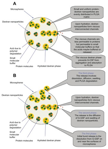
Figure 9 (A and B) The released mechanism diagram from the PLGA microsphere. (A) Mechanism diagram stability of G-CSF; (B) Released mechanism diagram of G-CSF.
Abbreviations: G-CSF, granulocyte colony–stimulating factor; PLGA, polylactic-co-glycolic acid.