Figures & data
Figure 1 Structures of deoxycytidine, cytosine arabinoside, and gemcitabine.Citation6
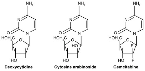
Figure 2 Schematic representation of a liposomal structure with a characteristic microenvironment and possible drug encapsulation as a function of its physicochemical features.
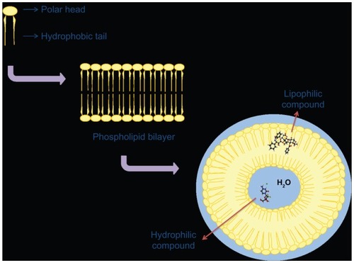
Figure 3 Schematic representation of the gemcitabine encapsulation process in liposomes using a pH gradient elicited by coencapsulation of a 250 mM ammonium sulfate solution. ©2006 Oxford University Press. Reproduced with permission from Celano M, Calvagno MG, Bulotta S, et al. Cytotoxic effects of gemcitabine-loaded liposomes in human anaplastic thyroid carcinoma cells. BMC Cancer. 2004;4:63.Citation35
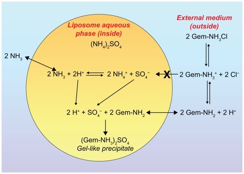
Figure 4 Chemical structure of PEG-folate-gemcitabine macromolecule and process for release of the nucleoside analog as a consequence of pH variation.
Abbreviation: PEG, poly(ethylene glycol).
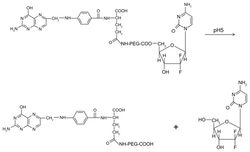
Figure 5 Dose-dependent cytotoxic effect of free gemcitabine (circle) versus gemcitabine-loaded PEGylated unilamellar liposomes (upwards triangle) against anaplastic thyroid carcinoma cells at different exposure times of 12 hours (A), 24 hours (B), 48 hours (C), and 72 hours (D).
Notes: The cytotoxic effect of the drug is expressed both as the percentage cell mortality (filled symbols and solid line) and the percentage cell viability (hollow symbols and dashed line). Cell mortality was evaluated by Trypan blue dye exclusion assay, while cell viability was evaluated by MTT testing. (■, □) represents untreated control cells and always shows mortality ≤ 5.5% and cell viability ≥ 97%. Unloaded liposomes showed similar values to controls (data not reported). Error bars, if not shown, are seen as symbols. Results are presented as the mean ± standard deviation of five different experiments. ©2008 American Scientific Publishers. Reproduced with permission from Celia C, Calvagno MG, Paolino D, et al. Improved in vitro anti-tumoral activity, intracellular uptake and apoptotic induction of gemcitabine-loaded pegylated unilamellar liposomes. J Nanosci Nanotechnol. 2008;8(4):2102–2113.Citation54
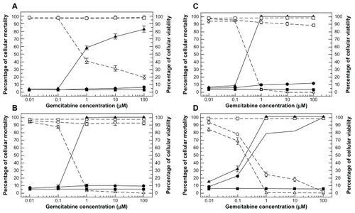
Figure 6 Confocal laser scanning micrographs of B-CPAP cells treated with fluorescein-labeled PEGylated unilamellar liposomes after 6 hours of incubation. (A) Hoechst filter, (B) FITC filter, (C) transmission mode, and (D) overlay.
Note: Bar 30 μm.Citation51
Abbreviations: FITC, fluorescein isothiocyanate; PEG, poly(ethylene glycol).
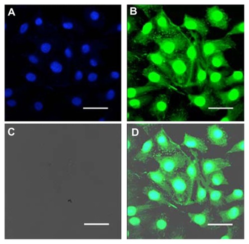
Figure 7 Flow cytometry cell cycle analysis of INA-6 cells. Control cells (A, D, and G), cells treated with free gemcitabine (B, E, and H), or with liposome-entrapped drug (C, F, and I) for 24 hours (A–C), 48 hours (D–F), or 72 hours (G–I). © 2008, Elsevier. Reproduced with permission from Celia C, Malara N, Terracciano R et al. Liposomal delivery improves the growth-inhibitory and apoptotic activity of low doses of gemcitabine in multiple myeloma cancer cells. Nanomedicine. 2008;4(2):155–166.Citation55
Notes: The symbol representing cells in S-phase is indicated; the symbol representing cells in sub-G1 phase is indicated as “apoptosis” because cells in the sub-G1 phase are recognized as being apoptotic.
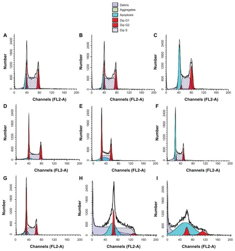
Figure 8 Interactions between gemcitabine-loaded PEGylated liposomes and pancreatic cancer cells. © 2011, Elsevier. Reproduced with permission from Yang F, Jin C, Jiang Y, et al. Liposome based delivery systems in pancreatic cancer treatment: from bench to bedside. Cancer Treat Rev. 2011;37(8):633–642.Citation25
Notes: Confocal laser scanning microscopy shows efficient interaction between gemcitabine-loaded PEGylated liposomes and BxPC-3 and PSN-1 cell membranes. Intracellular localization of fluorescein-dihexadecanoyl phosphoethanolamine is time-dependent.
Abbreviation: PEG, poly(ethylene glycol).
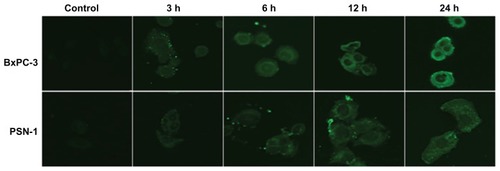
Figure 9 Examples of in vivo luciferase measurements, quantification, and generation of tumor growth curves, showing one animal from the control group (right) and one from the liposomal gemcitabine 8 mg/kg group (left). © 2007, Springer. Reproduced with permission from Bornmann C, Graeser R, Esser N, et al. A new liposomal formulation of gemcitabine is active in an orthotopic mouse model of pancreatic cancer accessible to bioluminescence imaging. Cancer Chemother Pharmacol. 2008;61(3):395–405.Citation56
Notes: Overlays of a picture with the light signal encoded as a spectrum with red representing the most, and blue the least intense light, are shown. Tumor end-volumes were 0.45 cm3 (1,122), and 1.69 cm3 (1,123), respectively.
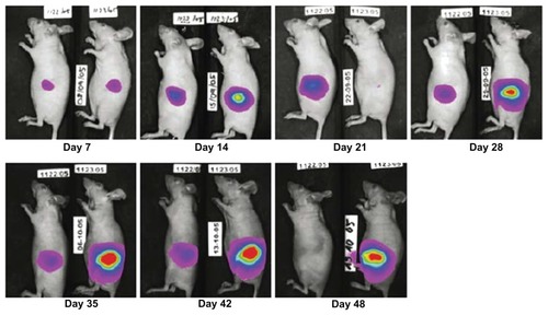
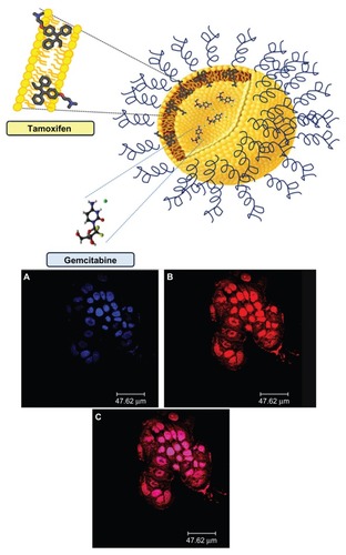
Figure 10 Schematic representation of gemcitabine-tamoxifen localization inside the multidrug carriers (upper panel). Confocal laser scanning micrographs of T47D cells treated with rhodamine-labeled PEGylated unilamellar liposomes after 6 hours of incubation. (A) Hoechst filter, (B) TRITC filter, and (C) overlay (lower panel).
Note: © 2012, Elsevier. Reproduced with permission from Cosco D, Paolino D, Cilurzo F, Casale F, Fresta M. Gemcitabine and tamoxifen-loaded liposomes as multidrug carriers for the treatment of breast cancer diseases. Int J Pharm. 2012; 422(1–2):229–237.Citation69
Abbreviation: PEG, poly(ethylene glycol).