Figures & data
Table 1 Characterization of Tested Nanocarriers
Figure 1 Biodistribution of core-shell polyelectrolyte nanocarriers and fluorescent products of their degradation.
Notes: BALB/c mice were injected with free Rhodamine B (RhB), poly-l-lysine fluorescently labeled with Rhodamine B (PLL-RhB) and PEG-terminated polyelectrolyte nanocarriers with fluorescently labeled shell (NC-RhB-PEG). Subsequently, animals were placed in the metabolic cages, and urine was collected. Mice were sacrificed 30 minutes after administration, and bile, serum, homogenates of liver, spleen, and kidney were obtained. The samples were analyzed for the fluorescence intensity at λ 560/590 nm. Bars represent the mean ± SD (n = 3).
Abbreviation: RFU/g, the parameter corresponding to fluorescence intensity measured for isolated fluid or tissue homogenate and calculated taking into account weight of isolated fluid or tissue.
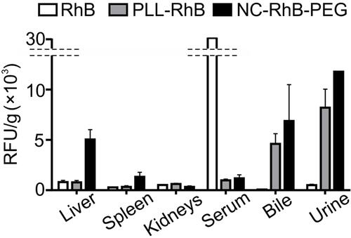
Figure 2 Biodistribution profiles and elimination routes of polyelectrolytes nanocarriers with the different core structure.
Notes: Tumor-bearing BALB/c mice were injected with PEG-terminated polyelectrolyte nanocarriers with liquid core (NC-RhB-PEG) or PEG-terminated polyelectrolyte nanocarriers with solid polycaprolactone core (PCL-RhB-PEG) with fluorescently labeled shell. (A) Mice were sacrificed 24 hours after administration, and serum, homogenates of liver, spleen, kidney, and tumor were obtained. The detected dose parameter was calculated (see Methods). Before the euthanasia, animals were then placed in metabolic cages, and (B) urine or (C) feces were collected over 24 hours. The samples were analyzed for the fluorescence intensity at λ 560/590 nm. Bars or points represent the mean ± SD (n = 3).
Abbreviation: RFU/g, the parameter corresponding to fluorescence intensity measured for isolated fluid or tissue homogenate and calculated taking into account weight of isolated fluid or tissue.
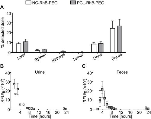
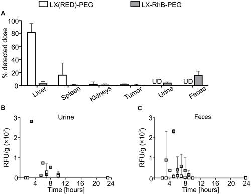
Figure 3 Biodistribution profiles and elimination routes of differently labeled latex-based nanocarriers.
Notes: Tumor-bearing BALB/c mice were injected with PEG-terminated polyelectrolyte latex nanocarriers fluorescently labeled in core (LX(RED)-PEG) or shell (LX-RhB-PEG). (A) Mice were sacrificed 24 hours after administration, and serum, homogenates of liver, spleen, kidney, and tumor were obtained. The detected dose parameter was calculated (see Methods). Before the euthanasia, animals were then placed in metabolic cages, and (B) urine or (C) feces were collected over 24 hours. The samples were analyzed for the fluorescence intensity at λ 560/590 nm. Bars or points represent the mean ± SD (n = 3).
Abbreviations: RFU/g, the parameter corresponding to fluorescence intensity measured for isolated fluid or tissue homogenate and calculated taking into account weight of isolated fluid or tissue; UD, under the detection limit.